Download
1 / 28
280 likes | 394 Views
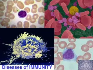
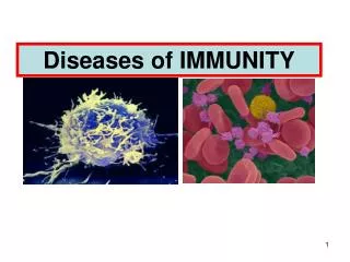
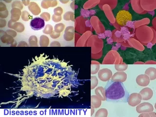
Diseases of Immunity (1). Hypersensitivity Reactions. Normally, a balanced system optimizes the eradication of infecting organisms without serious injury to host tissues.

E N D
Hypersensitivity Reactions • Normally, a balanced system optimizes the eradication of infecting organisms without serious injury to host tissues. • However, immune responses may be inadequately controlled or inappropriately targeted to host tissues, and in these situations, the normally beneficial response will cause of disease.
Hypersensitivity Reactions • The term hypersensitivity is used to describe immune responses which are damaging rather than helpful to the host. • WHY? • This term originated from the idea that individuals who mount immune responses against an antigen are said to be "sensitized" to that antigen, and therefore, pathologic or excessive reactions are manifestations of "hypersensitivity."
CAUSES OF HYPERSENSITIVITY REACTIONS • Autoimmunity. • Reactions against microbes. • Reactions against environmental antigens.
Hypersensitivity ReactionsClassification: • Type I “Allergy & anaphylaxis” • Type II “Antibody dependant” • Type III “Immune complex - mediated” • Type IV “Cell-mediated (delayed type)” • N.B: First 3 types are antibody-mediated injury & the last type is cell-mediated injury
Type I Hypersensitivity Reaction • ALLERGIC REACTION • ANAPHYLACTIC REACTION
Type I Hypersensitivity • Definition : • It is rapidly developing immunologic reaction occurring within minutes after the interaction of an antigen (allergen) with IgE antibodies bound to surface of mast cells or basophils in individuals previously sensitized to the antigen
Type I Hypersensitivity • Antigen is usually exogenous , environmental and is called allergen • Allergens may be introduced by inhalation, ingestion, touch, or by intravenous injection. • The reaction is mediated byIgE produced by previously sensitized B lymphocytes.. • IgE binds to the main effector cells ; the mastcell (in tissues) or basophils (in blood). • The initiated reaction passes through 2 phases an early one and a late one.
Type I Hypersensitivity:Phases • As Seen in the localized reactions • Initial response: • Characterized by; • Vasodilatation, • Vascular leakage, and • Smooth muscle spasm • Increased glandular secretions • These changes become evident within 5 to 30 minutes after exposure to an allergen and subside in 60 minute. • It is mainly mediated by histamine released by MAST CELLS.
Type I Hypersensitivity:Phases • Late-phase reaction: • Develop in 2 to 8 hours later without additional exposure to antigen and lasts for several days. • Characterized by intense infiltration of tissues with eosinophils, neutrophils, basophils, monocytes, and CD4+ T cells, and tissue destruction. • Occurs under effect of esinophils and neutrophils chemotactic factors released by mast cells.
Type I Hypersensitivity:Clinical Manifestations • The resultant reaction can occur as a systemic disorder or as a local reaction depending on the route of entry of allergen
Type I Hypersensitivityclinical presentation Local reactions: • Antigen is confined to a particular site • Skin contact Localized cutaneous swellings (urticaria, hives) and eczema, • Ingestion Allergic gastroenteritis (food allergy) diarrhoea • Inhalation bronchospasm allergic rhinitis, and bronchial asthma .
Type I Hypersensitivity:Clinical Manifestations • Systemic Anaphylaxis: • This usually follows an intravenous injection of an antigen to which the host has already became sensitized for. • systemic vasodilatation “anaphylactic shock” is produced and even death within minutes • Example: IV administration of Penicillin, Bee venom. • NOTE: The immune response in the late-phase inflammatory reaction, plays an important protective role in parasitic infections.
Type II Hypersensitivity reaction
Type II Hypersensitivity Antibody-mediated diseases; • Target antigens are present on the surface of cells or other tissue components • The antigens may be intrinsic to the cell membrane, or they may take the form of an exogenousantigen, such as a drug metabolite, adsorbed on the cell surface • The antibodies unite with the antigens MAINLY in the bloodstream, • This union sets off the complement system, and destruction of the local tissue cells ensues.
Type II HypersensitivityMechanism • A,Opsonization • B,Inflammation • C,Antireceptor antibodies
Type II HypersensitivityMechanism • Clinical examples; • Incompatible transfusion reactions (mismatched blood transfusion reaction) • Erythroblastosis fetalis “Rhesus antigen incompatibility” • Autoimmune haemolytic anemia, or thrombocytopenia • Certain drug reactions “penicillin hemolysis”
Type III Hypersensitivity reaction
Type IIIHypersensitivityImmune Complex - Mediated • Type III hypersensitivity is mediated by the deposition of antigen-antibody complexesformed in blood vessels. • The antigens may be • Exogenous antigens, such as bacteria, or viruses • Endogenous antigens, such as DNA. • Immune complexes deposit in bloodvessels in various tissue beds ,they have the ability to fix complement and trigger the subsequent injurious inflammatory reaction(either systemic or Localized )
Systemic Immune Complex DiseaseMechanism • Favoured sites of immune complex deposition are; • Renal glomeruli, • Joints, • Skin, • Heart, • Serosal surfaces, • Small blood vessels
Type IIIHypersensitivityExamples Autoimmune diseases as: • Systemic lupus erythematosis • Scleroderma • Sjogren syndrome
Type IV Hypersensitivity reaction
Type IVHypersensitivity(Cell Mediated) • Two types of T-cell reactions are capable of causing tissue injury and disease: • (1) delayed-type hypersensitivity (DTH), initiated by TH1-type CD4+ T cells • (2) direct cell cytotoxicity, mediated by cytotoxic CD8+ T cells that are responsible for tissue damage.
1-Delayed-Type Hypersensitivity • It is responsible for mediating immune reaction in case of; • Defence against variety of intracellular persistent or non-degradable antigens, such as tubercle bacilli. • pathogens, including mycobacteria, fungi, and certain parasites, • It may also be involved in transplantrejection. • Tumour immunity • NOTE: • In AIDS loss of CD4+ T lymphocytes increased susceptibility for developing TB & fungal infection.
Delayed-Type HypersensitivitySequence of Cellular Events • On subsequent exposure of an individual previously sensitised, the memory TH1 cells interact with the antigen on the surface of APC . • APC become activated, they produce IL-12 that activate CD+4 cells. • In turn CD+4 cells will secrete: • IFN-γ that activates macrophages to produce substances that cause tissue damage and promote fibrosis, • TNF promotes inflammation. • When macrophages become activated they transform into epithelioid cells And they occasionally fuse multinucleated giant cells.
Delayed-Type HypersensitivityMorphology • DTH is characterized histologically by the formation of Granuloma (a specific form of chronic inflammation) • It refers to microscopic aggregate of epithelioid cells, usually surrounded by a collar of lymphocytes with or without the formation of multinucleated giant cells.
Example for DTHTuberculin reaction • A classic example of DTH elicited by antigen challenge in an individual already sensitized to the tubercle bacillus by a previous infection. • Between 8 and 12 hours after intracutaneous injection of tuberculin (a protein extract of the tubercle bacillus), a local area of erythema and induration appears, reaching a peak (typically 1-2 cm in diameter) in 24 to 72 hours (hence the adjective, delayed) and thereafter slowly subsiding.
2- Direct cell cytotoxicity • In this variant of type IV hypersensitivity, sensitized CD8+ T cells kill antigen-bearing target cells • These effector cells are called cytotoxic T lymphocytes (CTLs) • They mediate their action through class I MHC molecule, where they directly lyse infected cells or stimulates their apoptosis. • It plays an important role in graft rejection, resistance to virus infections, and possibly tumor immunity