Download
1 / 1
10 likes | 95 Views
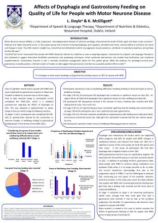
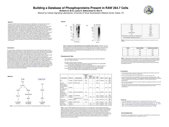
M. Ctrl. CLA. Ctrl. M. PVD. A. B. 250. 250. 150. 150. 100. 100. 75. 75. 50. 50. 37. 37. 25. 25. P-Thr-Pro. P-Tyr. Building a Database of Phosphoproteins Present in RAW 264.7 Cells Brekken D, Bi Q, Lyons K, Sethuraman D, Shu H

E N D
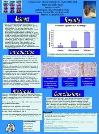
M Ctrl CLA Ctrl M PVD A. B. 250 250 150 150 100 100 75 75 50 50 37 37 25 25 P-Thr-Pro P-Tyr Building a Database of Phosphoproteins Present in RAW 264.7 Cells Brekken D, Bi Q, Lyons K, Sethuraman D, Shu H Alliance for Cellular Signaling Laboratories, University of Texas Southwestern Medical Center, Dallas, TX Results Abstract A goal of the AfCS Protein Chemistry Laboratory is the identification of phosphoproteins present in the AfCS model cell systems. This poster describes the identification of a large number of phosphoproteins and phosphorylation sites in the RAW 264.7 macrophage cell line. RAW cells were treated with either tyrosine or serine/threonine phosphatase inhibitors to generate high levels of protein phosphorylation. Phosphoproteins and phosphopeptides were enriched using immuno-affinity chromatography with anti-phosphotyrosine antibodies and immobilized metal affinity chromatography (IMAC), either alone or in combination. Phosphoproteins and phosphorylation sites were then identified by tandem mass spectrometry. A total of 248 proteins and 210 distinct serine, threonine, and tyrosine phosphorylation sites were identified. The proteins identified included integral membrane proteins and proteins likely to be present at low levels in these cells. Phosphoproteins identified included kinases, phosphatases, transmembrane receptors, signaling proteins, cytoskeletal proteins, translation factors, cell adhesion proteins, and proteins involved in RNA metabolism. Many of the known proteins identified are components of macrophage signaling pathways. The list of proteins also includes a large number of proteins not previously known to be phosphorylated as well as completely novel proteins. Table 2. A table listing a sample of identified proteins that are known to be involved in immune signaling. Figure 2. Generation of phosphorylated proteins by phosphatase inhibitor treatment. RAW 264.7 cells were either left untreated (Ctrl), treated with 25 nM CLA (CLA), or treated with 100 uM PVD (PVD) for 30 min and lysed in SDS sample buffer. The proteins were resolved on a 10% SDS gel, transferred to nitrocellulose membranes, and immunoblotted with either an anti-phospho-threonine-proline antibody (P-Thr-Pro-101) (Panel A) or an anti-phosphotyrosine antibody (P-Tyr-100) (Panel B) from Cell Signaling Technology. The mass of molecular weight markers (M) are shown at the left. Introduction An important goal of the Alliance for Cellular Signaling (AfCS) is the global analysis of ligand-induced changes in protein phosphorylation. Important steps in this process are the identification of the repertoire phosphoproteins present in the AfCS model cell systems and determining their sites of phosphorylation. Due to the low abundance of many phosphorylated proteins and the fact that they are often phosphorylated at low stoichiometries, an enrichment step is usually needed in order to identify a broad spectrum of phosphorylated proteins in a given cell type. One useful method for enrichment of phosphoproteins is immuno-affinity isolation with antibodies that recognize phosphorylated amino acids. Anti-phosphotyrosine antibodies are particularly useful in this regard (Shu et al., 2003). Enrichment of phosphopeptides with immobilized metal affinity chromatography (IMAC) has allowed the identification of hundreds of phosphoproteins and phosphorylation sites in yeast (Ficarro et al., 2002), Arabidopsis plasma membrane proteins (Nuhse et al., 2003), and in WEHI 231 B lymphoma cells (Shu et al., 2004) . We have used a combination of these approaches to identify phosphoproteins and their phosphorylation sites in the murine RAW 264.7 macrophage cell line following treatment with phosphatase inhibitors. The list of proteins and phosphorylation sites is presented as a supplement to this poster. Because this is an ongoing project, the table will be updated periodically and eventually incorporated into a database directly accessible through the AfCS website. Phosphoprotein Table Table 3. Phosphorylation sites identified and activation loop residues in several proteins identified. The tyrosine phosphorylation sites identified in Fgr is a known sites within the activation loop that is associated with activation of kinase activity. In contrast, the phosphorylation sites identified in Btk, Lck, and Lyn were all threonine residues immediately adjacent to the activating tyrosine phosphorylation site of these kinases. These threonine phosphorylation sites were all identified in pervanadate treated cells. The complete table (available upon request) lists the phosphoproteins identified in RAW 264.7 cells from multiple experiments. The table contains 3 classes of proteins: 1) proteins identified following SDS electrophoresis of anti-phosphotyrosine immunoprecipitates (PY ID) where a phosphorylation site was not identified; 2) proteins identified following IMAC enrichment of phospho-peptides from whole cell lysates of calyculin A-treated cells (IMAC) where phosphorylation site(s) were identified; and 3) proteins that were identified following anti-phosphotyrosine immunoisolation and IMAC enrichment (PY IMAC) where a phosphorylation site was identified. • Conclusions • The phosphoprotein table contains 481 separate protein identifications that identify a total of 248 proteins from three different experimental approaches. • 210 unique phosphorylation sites were identified (59 P-Tyr, 116 P-Ser, and 35 P-Thr). • A total of 24 novel proteins were also identified. These are listed as “hypothetical proteins”, “Riken cDNAs”, or “Unknown protein products”. The identification of peptides corresponding to these proteins not only established that they are expressed in RAW 264.7 cells, but also shows they are phosphorylated. • A large number of proteins known to be involved in signaling in macrophages and other immune cells were also identified (Table 2). • This information will be used to expand the signaling “parts list” identified in these cells and allow the development of probes that will be used to monitor ligand-dependent changes in protein phosphorylation. Methods P-Tyr P-Ser/P-Thr PVD CLA P-Tyr IP Trypsin 1D gel Trypsin Esterification Trypsin IMAC IMAC References • Ficarro SB, McCleland ML, Stukenberg PT, et al. (2002) Nat. Biotechnol. 20(3), 301-305. • Nuhse TS, Stensballe A, Jensen ON, and Peck SC (2003) Mol. Cell Proteomics 2(11), 1234-1243. • Shu H., Chen S, Lyons K, Hsueh R, Brekken D (2003) AfCS Research Reports. Vol. 1, no. 3 [cited March 21, 2003]. Available from: http://www.signaling-gateway.org/reports/v1/DA0003.pdf • Shu H, Chen S, Bi Q, Mumby M, Brekken DL (2004) Mol. Cell Proteomics 3(3), 279-286. LC-MS/MS (PY ID) LC-MS/MS (PY IMAC) LC-MS/MS (IMAC) Figure 1. Flow chart of procedure for the enrichment of phosphorylated proteins from whole-cell lysate. Table 1. Sample of RAW Phosphoprotein table. The table lists the name and protein GI number that was identified. Where possible, the protein name is derived from the NCBI LocusLink entry. If a phosphorylation site was identified, the phosphopeptide detected is listed with an asterisk (*) following the phosphorylated amino acid. There is a column that lists the residue number of the identified phosphorylation site(s) within the amino acid sequence of the protein. Note that these numbers are based on the sequence listed in the corresponding GI entry and do not always represent the full-length sequence. For proteins that were identified directly by LC-MS/MS (i.e., without an IMAC step), the highest Mascot score and # of peptides detected are listed. The Mascot score is the absolute probability that the observed match is a random event (-10*Log(P), where P is the probability that the observed match is a random event). Mascot scores greater than 62 were considered significant (p<0.05). If available, the LocusLink and AfCS ID numbers are also listed in the table. The table also includes columns listing the number and type of experiment where the protein was identified. Acknowledgements We would like to acknowledge Robert Cox, Laura Draper, Farah El Mazouni, and Marc Mumby from the AfCS Protein Chemistry lab for their contributions to this work.