Download
1 / 80
930 likes | 1.33k Views
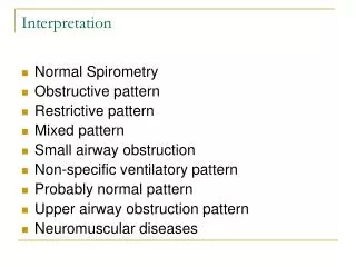
Laboratory Interpretation. K.Thornton MSN,RN. Blood Loss. Acute blood loss. Hemolytic anemia. Infection. Acute Infection. Sepsis. Coagulation Tests. HIT. DIC. Nutrition Function. Electrolytes. Sodium. Potassium. Magnesium. Rhabdomyolysis. Drug Monitoring. Lecture Outline.

E N D
Laboratory Interpretation K.Thornton MSN,RN
Blood Loss. Acute blood loss. Hemolytic anemia. Infection. Acute Infection. Sepsis. Coagulation Tests. HIT. DIC. Nutrition Function. Electrolytes. Sodium. Potassium. Magnesium. Rhabdomyolysis. Drug Monitoring. Lecture Outline
Update, 4:55 Blood Components • Plasma: 55% of whole blood • Erythrocytes: 44% of whole blood • Leukocytes & Platelets: <1% of whole blood 5:42
Hemoglobin (Hb): O2 binds to “heme” of Hb. Each RBC contains ~ 300 million molecules of Hb. Combines with 1.39 cc O2. 1 unit PRBC: incr hb by 1gm. Hematocrit (Hct): Percentage of RBC in whole blood. Hct is usually 3X Hb value. 1 unit PRBC: incr Hct by 3-4% Hemoglobin & Hematocrit 7:10
Acute Blood Loss • Manifestations are related to loss of blood volume rather than loss of hemoglobin. • Blood Volume = plasma + cells • Blood Loss: Loss of both plasma & cells. • Decrease in Hemoglobin: • reflects loss of cells only • Decrease in Hematocrit: • reflects loss of plasma & cells • (Example: Pt has 8hb & 24RBC, then receives 1unit. Now pt has 9hb & 27RBC)
Acute Blood Loss Plasma is replaced in a few hours Platelet count Internal Hemorrhage: Neutrophil count RBC. Hb. Hct. Iron recovered External Hemorrhage: Shift from tissues & interstitium to vascular Presence of myelocytes, metamyelocytes & nucleated RBC in circulation Iron is depleted Shift of marginated WBC into circulation Incr Erythropoiesis
Hemolytic Anemia • Premature, accelerated destruction of RBCs. • Accelerated Erythropoiesis (8xnormal): • BM’s “bone marrow” response to RBC destruction. • CBC: “Complete blood count” • Decr RBC count, Hct, & Hb • RDW:(Incr RBC Distribution Width- “the size of RBC”) • b/c of Anemia, RBC loss, and sickle cell disease • Increased in variation
RBC Destruction & Bilirubin Spleen RBC Hemoglobin Liver Globin Heme Glucuronide Indirect Bilirubin (Unconjugated) Direct Bilirubin (Conjugated) Kidney Bacteria Duodenum Urobilinogen Urobilinogen Stool Urine
Hemolytic Anemia • Reticulocyte Count: immature cellsincreased • Causes: Sickle cells &/or alcoholism • s • Indirect (Unconjugated Bilirubin): increased • An early sign of liver disease/problems • Urine Urobilinogen: increased • An early sign of liver disease • Serum Haptoglobin (Hp): decreased • Meaning you have sickle cell & Thaslassemia • Sign of bloodloss: 1st see change in consciousness & restlessness. THEN, H&H will be affected/show.
Acute Infection / Inflammation / Injury ESR Albumin C-Reactive Protein (CRP) Increases Elevated means infection or injury Decreases A protein, body Is using it up for Energy. hs-CRP: Assess risk of MI
Viral Infections Decreased WBC Count Increased Lymphocytes Its just the nature of the infection…
Acute Bacterial Infection / Inflammation / Injury WBC Count Recovery: Acute: Sepsis: Towards normal values Will see Decrease b/c you’re screwed Will see increase Increased Monocytes (Phagocytes) = recovery phase of acute infections
Acute Infection/Inflammation/ Injury Neutrophil Count 3000 – 7000 /mm3 or ~ 45 - 75% of total WBC Count. Sepsis: Decreased Acute Bacterial: Increased Decr Segs Will see Incr Bands Incr Segs Recovery: Incr bands Metamyelocytes & Myelocytes Decr Bads. Incr Segs. 0 metas & myelos
Shifts in WBCsa shift of cells. Regenerative means you’re getting better. 0 Bone Marrow Response • Degenerative Shift to the Left: • Increase in band cells with no leukocytosis. • Poor prognosis (Degenerative = Down) • Regenerative Shift to the Left: • Increased band cells with leukocytosis. • Good prognosis • Shift to the Right: (Right recovery) • Decreased band cells with increased segmented neutrophils. Bone Marrow Response
Response to Infection & Injury Local Inflammatory Response Systemic Inflammatory Response (SIRS). When SIRS is caused by microorganisms Sepsis Sepsis w/ hypoperfusion & organ dysfunction Severe Sepsis Sepsis w/ hypotension despite fluid resuscitation Septic Shock 39:18
Septic ShockFluid is the primary treatment for sepsis. • Criteria:(Any 3) • Temperature < 36 oC (96.8F)OR > 38 oC (100.4F). • HR > 90 bpm. • RR > 20 /min OR PaCO2 < 32 mm Hg. • WBC count < 4,000 cells/mm3, OR > 12,000 cells/mm3, OR > 10% band forms. • Lactate level 4 or greater = sepsis • ~ 750,000 new cases/year & 200,000 deaths. • Estimated annual cost of Rx > $16 Billion!
Bacteria. Trauma. Shock Sepsis Pathophysiology Inflammatory Response. Release of chemical mediators. Endothelial damage Vascular permeability Stimulation of coagulation. DIC. Activated Protein C Vasodilation. Peripheral edema. Hypotension. Tissue damage. Organ failure. Decreased immune function. Clinical Manifestations: Tachypnea. Tachycardia. Fever. Leukocytosis, followed by leukopenia.
Later Sepsis: Hypodynamic CO & SVR Lactic acid Tissue /organ damage cellular oxygenation Coagulation cascade Early Sepsis: Hyperdynamic CO & SVR DIC Perfusion Imbalance Hematological Alteration Myocardial Alteration Dilated Ventricles SEPSIS Broncho- constriction Hyperglycemia 2o insulin resistance Pulmonary Alteration Metabolic Alteration Interstitial & alveolar edema Impaired gas exchange serum amino acids. serum ketones. Lactic acid. Tissue & organ death ARDS
Perfusion Imbalance Inflammatory mediators & Bacterial endotoxins Activation of coagulation cascade Vasodilation & increased capillary permeability Microthrombi in capillaries Activation of Sympathetic Nervous system Low SVR & hypovolemia Tissue hypoxia & organ damage Vasoconstriction of splanchnic circulation & vessels of the skin with dilation of skeletal muscle beds Inadequate cellular Oxygenation (Inflam response) Increased lactic acid production Anaerobic metabolism Blood flow only to vital organs
Myocardial Alterations High CO & Low SVR Hyperdynamic Heart Early Sepsis Initial mechanism leading to heart failure! Low CO & Increased SVR Late Sepsis Hypodynamic Heart MYOCARDIAL DEPRESSION + Lactic Acidosis Circulating Depressants Dilated cardiomyopathy
Pulmonary Alterations Inflammatory Chemical Mediators & Bacterial Endotoxins BRONCHOCONSTRICTION Increased work of breathing Increased capillary permeability Pulmonary Hypertension Interstitial edema Alveolar edema Shunting Medium for bacterial growth Impaired gas exchange ARDS Hypoxemia
Metabolic Alterations Excessive Catecholamines “Your body’s natural Adrenaline” Stimulates gluconeogenesis Hyperglycemia Glycogen stores are depleted Insulin resistance Cells unable to use glucose, protein & fat Protein used for energy Fats used for energy Increased ketones Increased serum amino acids Cellular pumps fail Lactate production TISSUE & ORGAN DEATH
Hematological Alteration: Coagulation Cascade in Septic Shock APC Micro- organism Activation of Coagulation Inactivation Endotoxin Release Tissue Factor Factor VIIa Factor Va Stimulates Thrombin activation Inhibits Inhibits Fibrin clot formation Fibrinolysis APC Depletion of clotting factors DIC Tissue Factor Endothelial cells Hemorrhage
Sepsis: Labs • Cultures: From wound, UA, Catheter • CBC: Will see an incr or decr in WBC (so look for the words early or late) • Bands will incr, meaning sepsis. • Chemistry: will develop into hyperglycemia. • Steroids may be given which is another reason for insulin drip. • ABG: Metabolic acidosis b/c of impaired gas exchange. • Lactate Levels (Normal 0.5-2.0): Will incr & incr SVR. 54:50
Sepsis: Labs Continued…Note: Xigris is a protein replacement. Has anti-thrombotic, anti-inflammatory, and profibrinolytic properties. To prevent DIC & to treat sepsis. • Hemodynamic Monitoring: Fluids to treat hypovolemia. • Decr CardOutput and incr SVR • CXR & Abdominal X-rays: • infiltrates • CT Scans: • Abscesses and anything that can be infectious or cause an infection. 1:01:10, break
COAGULATION CASCADE- what happens to make a clot Intrinsic Pathway Extrinsic Pathway XII XIIa VIIa VII XI XIa Tissue Factor IX IXa X Xa X XIII V Va Prothrombin Thrombin XIIIa Fibrinogen Soluble Fibrin Insoluble Fibrin
Platelet CountLife span of platelet is 7.5days • Normal Counts: 140 - 400 x 103/mm3. • 2/3 ( circulating in blood ) & 1/3 ( spleen ). • Drugs Affecting Platelet Function: • Plavix, aspirin, Coumadin & NSAID (ibuprofen) • Thrombocytosis: • increased risk of clot • Seen in CA, splenectomy, chronic pancreatitis • Thrombocytopenia: • increased risk of bleeding 1:07:30
Bleeding TimeMeasures the primary stage of hemostasis. • Phase: Interaction of platelet with blood vessel wall & formation of hemostatic plug. • Best screening test for platelet function disorders. • Prolonged: means thrombocytopenia, no clotting factor, leukemia, and/or DIC. • Common meds for surgery: • Protonix- To reduce gastric acid. A prophylaxis to protect GI Tract. Since they’re taking a blood thinner. Its to protect from stomach ulcers. • Lovenox- to prevent & treat deep vein thrombosis or pulmonary embolism.
HIT- Heparin Induced Thrombocytopenia • HIT Type II or White Clot Syndrome: • Heparin-dependent antibodies develop after a patient has been on heparin for 5 or > days or previous heparin exposure. • Presence of Factor 4 can induce Heparin Induced Thrombocytopenia. • Normal person • Non-immune Heparin Associated Thrombocytopenia (non-immune HAT): • Absence of heparin-dependent antibodies. • HIT Type I. • Not common
HIT: Pathophysiology of Heparin-PF4 complex formation Antibody generated against Heparin-PF4 complex “Antibody-Heparin-PF4” attaches to platelets Activation of platelets releases microparticles (procoagulant) Activates coagulation cascade
HIT: Diagnosis • Normal platelet count before commencement of heparin therapy. • Onset of thrombocytopenia 5 - 14 days after initiation of heparin therapy. (“hmm, works too well”) • Platelets drop, so then start to consider Lovenox. • R/O other causes of thrombocytopenia. • Occurrence of thromboemboli during heparin therapy. (Here nurse says, “this aint right!?!”) • Platelet aggregation assay/panel. Common, cheaper. • Serotonin release assay/panel. Definitive test, but expensive.
HIT: Treatment • Avoid use of heparin. • Heparin drips (IV). • Lovenox (SQ). • Heparin flushes. • Heparinized dialysate. • Warfarin (???) a: no. • Anticoagulants. • ARGATROBAN. • Advantage: • Monitor: Thrombosis. Platelet counts. Venous Limb Gangrene Would the physician order platelet transfusions? 1:17:18
Prothrombin Time (PT) • Protein produced in the liver – depends on Vitamin K intake & absorption. • Results are reported as: • Time (seconds): Normal 12-15 seconds • INR:2.0 to 3.5 • Effectiveness of Coumadin Therapy. • ***Maintain PT 2 to 2.5 times normal range. • Prolonged: will bleed • Shortened: will form clots • Antidote: Vitamin K. NovoSeven. ($7,800-10,000/dose)
Partial Thromboplastin Time (PTT) • Normal PTT: 60-70 seconds (Varies!) • Activated Partial Thromboplastin Time (APTT) • Normal: 21-35 seconds • Prolonged PTT: • You will see this w/ heparin treatment, decr of Vitamin K, Liver disease, DIC • Shortened PTT: • Immediately after an acute hemorrhage, and/or Early DIC
Heparin Therapy • When rapid anticoagulation is desired. • Monitor: • PTT: 1.5 to 2.5 times normal. • Desired therapeutic range: 40-70 seconds • Too much heparin: If > 90 seconds • aPTT: 1.5 to 2.5 times normal. • Desired therapeutic range: 70 seconds • Too much heparin: > 100 seconds • Antidote: Protamine Sulfate. Given slowly.
IV Heparin Guidelines • Baseline Wt (kg), PTT, INR, CBC with Platelets. • Heparin: 25,000 units in 500 ml D5W • Concentration: • Bolus: 80 units/kg. • Maximum Bolus: • Infusion: 18 units/hg/hr • Maximum Rate: • Lab: • PTT (CHS: 24.1 – 37.3 sec) every 6 hours x 48 hours. • If 2 consecutive PTT in therapeutic range after 1 day PTT daily. • H & H daily. • Platelet count every 24 hours.
Fibrinogen: Converted to fibrin. Fibrin + Platelets → Clot. in injury & inflammation. Normal: 200-400mg/dL Decreased: Liver disease & DIC Panic value: Cryoprecipitate >700 mg/dL: MI/CVA risk D-Dimers: To check if pt is going into DIC. Produced by action of plasmin on fibrin. (Will see an incr in fibrinogen) Normal: < 250 ug/L Increased: Renal/Liver failure, sepsis. Fibrinogen & D-Dimers 1:29:26
Fibrin Degradation Products (FDP): Used to dissolve clots. Fibrin is split by plasmin → fibrin degradation (split) products. Normal: 0 or <10mg/L Critical value: > 40mg/L Increased: Primary: fibrinolysis Protein C: Produced by the liver. Prevents thrombosis (anticoagulant). Enhances fibrinolysis. Decreased: Means you have DIC, Liver disease, Vit K deficiency, ARDS, & CA FDPs & Protein C Thrombin Plasminogen Plasmin Fibrin FDP
DIC • Inappropriate triggering of the coagulation cascade & breakdown in normal feedback mechanisms in body that allow for distribution of clots. • Common causes: • Surgery. Obstetric emergencies. Liver cirrhosis, Trauma/crushing injs,. Transfusion reactions. Burns. Septicemia. Shock or low flow states.
DIC Pathology- 1:36:20 • Exessive bleed shock death. 1:37:30
DIC: Assessment • S/S of inappropriate clotting: • Thrombosis • Gangrene • Altered LOC. CVA • PE • Bowel ischemia & infarction • ARF • S/S bleeding: • Bleeding from various sites • Hematuria • Petechial reashes • Oozing from IV acess sites 1:40:00
DIC: Labs • Massive Intravascular Clotting & Secondary Depletion of Essential Clotting Factors: Platelet Count Decreased Fibrinogen Level Decreased Prothrombin Time (PT) Prolonged Early- Shortened Partial Thromboplastin Time (PTT) Late- Prolonged Protein C Level Decreased
DIC: Labs • Excessive / Accelerated Fibrinolysis: Fibrin Degradation Products (FDPs) increased increased D-dimer Assays decreased Antithrombin III Level
DIC: Labs • Clinical effects of microvascular clotting / cell destruction: • Schistocytes in peripheral smear: Present • Serum Bilirubin: increase b/c of the fibrolysis & hemolysis • BUN: incr b/c of the fibrolysis & hemolysis
Treat underlying cause. Correct Effects: Oxygenation (ischemia) replace fluids Electrolyte balance Vasopressors In Severe Bleeding: cryoprecipitate. Fresh frozen plasma platelet transfusions Heparin Infusion: Interrupts thrombosis process. Controversial. DIC: Treatment 1:27:40 Case study- 1:51:40
Metabolic Cart-b/c of everything incr calorie expendature. • Calculates REE (Resting Energy Expenditure). • Exact calorie consumption. • Temp incr of 1degreeF, - incr calorie demand 500kcal/day.
Prealbumin- to reflect status TODAY. • Short Half-life: 2-3 days • Responds QUICKLY to decreased nutritional intake & nutrition restoration. • Normal: 19-38mg/dL • Severe protein depletion: 0-5 • Moderate protein depletion: 5-10 • Mild protein depletion: 10-15
Urine Urea Nitrogen • 24-hour Urine Collection. • Measures degree of catabolism. • The more protein catabolized = the more urea present in the urine. • Normal: 5gm/24hrs • Severe:above 18gm/24hrs • Pts w/ bad Kidneys/Renal, this won’t work.