Download
1 / 32

E N D
Introduction • Radiography is a highly technical field, indispensable to the modern dental practice, but presenting many potential hazards. The dental radiographic specialist must be thoroughly familiar with the procedures necessary to produce radiographs of diagnostic quality. • He must also have a thorough knowledge of the hazards associated with the use of radiation and how to protect himself and the patient against those hazards. This lesson deals with the production, characteristics, and effects of radiation and how it may be used safely in dentistry.
Discovery • In 1895, Wilhelm Konrad Roentgen was searching for invisible light by experimenting with a Crookes vacuum discharge tube. This is a glass tube in which the vacuum is nearly complete, having a negative electrode (cathode) and a positive electrode (anode). Many investigators believed that invisible light rays were emitted from the negative electrode when a high voltage current was sent through the tube. With the room darkened and the tube covered with black paper, • Roentgen passed a high voltage current through the Crookes tube and was surprised to observe that a fluorescent screen lying on a table at some distance was glowing brightly. He then noted that a shadow was produced when an object was placed between the tube and the screen. Further experimentation revealed that the rays that caused the fluorescent screen to glow also acted upon the emulsion on photographic plates in the same manner as light. Thus it was shown that the rays produced would pass through some substances through which light would not pass. In later years scientists have referred to them as Roentgen rays.
Terms • Radiograph. An exposed and processed film (roentgenograph, roentgenogram). Also known as an x-ray negative. • Roentgenology. The study and use of x-rays (radiology). • Roentgen Ray. Electromagnetic radiation of pure energy and extremely short wavelength (X-ray), sometimes referred to as x-ray photons. • X-ray Photon. Electromagnetic rays produced by the x-ray machine.
Radiation • General. There are two sources of radiation (natural background radiation and man- made) both of which are harmful to man. • Natural Background Radiation. There are three sources of natural background radiation: cosmic, earth, and internal. Although natural background radiation may be harmful, man has lived in this environment without significant injurious effects since his appearance on earth. • Man-Made Radiation. Man-made radiation has many sources. Some of them are from medical and dental radiographs, occupational exposure, fallout from weapons testing, television sets, and certain radioactive watch dials, clocks, and meters. Man-made radiation, used improperly, can be significantly more harmful to man than natural background.
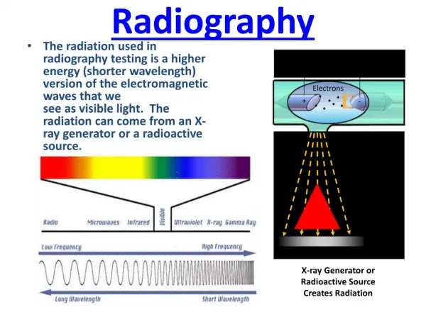
Types of Radiation • Particulate. Particulate or corpuscular radiation comes from radioactive decay or disintegration of radioactive materials. Alpha and beta particles are examples of this type radiation. • Electromagnetic. Electromagnetic radiation covers a very wide spectrum ranging from electrical power to visible light to x and gamma rays. The portion of the electromagnetic spectrum most important to us in this particular study is the x-ray portion.
Parts of an X-Ray Machine • General. The standard structural parts of the dental x-ray machine include a control panel (usually mounted behind a protective shield); a tube head, which houses the dental x-ray tube; and a flexible extension arm from which the tube head is suspended (see figure 1-1). • The Control Panel. The components of the control panel are switches, dials, gauges, and lights. Basically, each control panel has the same function, the arrangement and location of these components will differ, depending upon the make, model, and year of construction of the dental x-ray unit. An operator's manual is issued with each unit. The operator should study it until he is familiar with its operational capability. • The Extension Arm. The tube head is attached to the metal extension arm by means of a yoke that can revolve 360 degrees horizontally where it is connected. The construction of the yoke also provides vertical movement as well. • The Tube Head. Inside the metal tube housing is the x-ray tube. The diagram in figure 1-2 represents a dental x-ray tube head and a dental x-ray tube. This tube emits radiation in the form of photons (photons will be discussed in Lesson 2) or x-rays. X-ray photons expose the film. In addition to exposing the film, it also exposes the patient to radiation. Unless certain protective measures are taken, the x-ray technician may also be exposed.
Three Steps in X-Ray Production • The First Step. The first step in x-ray production is to turn on the machine. (If there is doubt on the part of the x-ray technician concerning the operation of the unit, reference should be made to the operator's manual.) When the unit is turned on, the filament of the cathode is heated by electrical current, causing it to emit electrons (see figure 1-3). • The Second Step. For the second step of this three-step process, high voltage is passed across the x-ray tube. When this is done, the electrons or electron cloud from the filament are drawn across the opening toward the anode. The anode is made of tungsten and is sometimes called the tungsten target. Figure 1-4 depicts the electrons speeding toward the anode (tungsten target). • The Third Step. The third and final step in this three-step process is the collision of electrons with the anode (tungsten target). This rapid deceleration of electrons produces x-rays, also referred to as photons. Figure 1-5 represents electrons striking the anode (tungsten target) and producing x-ray photons.
Hazards • The dental x-ray technician should never receive primary radiation from a dental x-ray unit if safety precautions are observed. However, scattered and/or secondary radiation is more difficult to avoid and is a serious danger to the technician. This type of radiation is produced by a scattering of the primary x-ray beam. • The x-ray photons and photo-electrons in the beam undergo a change of direction after interaction with atoms and molecules as they pass through a substance.
Radiation Protection • General. Filtration and collimation of the x-ray beam are very important safety measures. The filter and collimator (diaphragm) block the majority of the unwanted x-ray photons. As you progress through the next few paragraphs of this text, you will understand their importance. The following diagram will identify the location of these two devices • Filter. The aluminum filter or disk is placed in the path of the x-ray beam. It is located at the base of the cone or position indicating device (PID) just inside the metal housing. Figure 1-8 shows the location of the PID. The filter completely covers the opening where the x-ray beam emerges from the x-ray tube. The reason for the aluminum filter is to absorb the low energy, long wavelength x-rays (photons) and allow the high energy, short wavelength x-rays (photons) to pass through the filter. Filters on dental x-ray machines with over 70 kVp have a minimum thickness of 2.5 mm of aluminum. Those machines below 70 kVp have a safety standard minimum of 1.5 mm aluminum.
Radiation Protection Continued • Collimator. The lead diaphragm is collocated with the aluminum filter. It restricts the x-ray beam to the desired size. The diaphragm or collimator is constructed of 1/16-inch lead. • Without this collimator, x-ray photons would cover a wide area of the patient's head. With the lead diaphragm or collimator in place, only the area necessary for exposure receives the primary beam.
Protection Standards • (1) Radiation monitoring. AR 40-14 prescribes monitoring practices for Army personnel. It requires all primary x-ray technicians to wear a dosimeter or film badge. The dosimeter monitors or measures radiation received by the technician. The results are recorded on DD Form 1141. This form is kept permanently and made a part of the individual's health record. (2) Radiation standards. For the technician operating a dental x-ray machine, the level of radiation must not ever exceed an accumulated whole body dose, in rems, of five times the number of years beyond age 18 and a maximum of three rems in any 3-month period. NOTE: The term rem refers to "roentgen equivalent in man," a unit measuring the biological effect of radiation energy. For x-rays, 1 rem is equal to 1 rad, or "radiation absorbed dose" (rad). • (3) Protective booth and shields. Standards for dental x-ray booths or rooms require a shielding thickness of 1/16-inch lead or equivalent. The timer switch used to activate the machine for exposures is permanently affixed to the outside wall. The timer switch is mounted outside the protective shielding to prevent the operator from standing inside the booth during exposures. The shield is so designed that the radiation must scatter at least twice before reaching the x-ray technician. Leaded glass on the booth or shield provides a continuous view of the patient during the exposure. Consequently, any holding of the film or tube head by the x-ray technician is strictly prohibited.
Continued • Patient Protection. It is the responsibility of the x-ray technician to use all available protective measures to reduce exposure to the patient. Only those radiographs requested by the dental officer will be taken. Be sure that a good quality x-ray is produced each time a request is made. Wrong exposures, improper exposures, and faulty processing techniques must be avoided. These mistakes result in retakes and unnecessary patient exposure. Also, the lead apron must be used for every exposure. These safety devices significantly reduce patient exposure.
Quantity and Quality • The quality of the x-ray beam is controlled by the voltage, while the milliamperes control the quantity. An increase in the voltage and milliamperes reduces exposure time for the patient. • X-ray Beam Quality.Voltage provides contrast to the film. The desired contrast appears as various shades of gray, black and white in the x-ray negative (radiograph). Increased voltage provides less contrast (or more shades of gray). However, the beam has more penetrating power. Decreased voltage, on the other hand, provides more contrast (fewer shades of gray and more black and white shades). However, there is less penetrating power in the low voltage exposure. The technique most commonly used to expose periapical and bite-wing X-rays is a 90 kilovolt peak and 15 milliamperes. • X-ray Beam Quantity. The more x-rays (photons) in the x-ray beam, the more dense (dark) the x-ray negative (radiograph) becomes. By increasing the milliamperes, we increase the number of available electrons at the cathode filament. When electrical current (voltage) is applied to the x-ray tube, the electrons cross the gap. When they impact on the anode (tungsten target), a greater number of x-rays (photons) are also produced. The more x-rays that are available to penetrate an object, the more dense (dark) is the x-ray negative (radiograph).
X-Ray Photons • X-ray photons are electromagnetic rays produced in the x-ray tube head when electrons from the cathode filament strike the anode target. They are bundles of pure energy. The photons transfer their energy to the substance through which they pass whether it be air, an x-ray film, or the living tissue of the patient or the specialist. They cannot be seen or felt.
Photon Action Upon Atoms. • (1) Photon collision with the nucleus of an atom. The photon may strike the nucleus of an atom. If this occurs, the atom will be destroyed and the photon will release or expend its energy. See figure.
(2) A direct photon hit upon an electron by a photon. The photon may strike an electron with a direct hit. This action will result in the release of the photon's energy, transferring its energy to the electron. The electron will be dislodged from its shell. When an electron is dislodged in this manner, it is called a photo-electron. The dislodged or departing electron (now a photo-electron) will have energy to ionize or strike other electrons. This is a form of scattered/secondary radiation, as noted earlier in the text.
(3) An indirect photon hit upon an electron. The photon may strike one of the orbiting electrons with a glancing blow, dislodging the electron from its shell. By striking a glancing blow, the photon will still possess energy and go on to strike other electrons. The dislodged electron becomes a photon-electron and will have energy itself. It, too, may strike and dislodge other electrons. This is also a form of scattered/secondary radiation.
Photon Action • (1) Living cells are composed of atoms and molecules. If the structure of the atoms and molecules is changed, this may adversely affect the cell. When cells are exposed to ionizing radiation, the structure of some of the atoms and molecules within the cell are changed. • (2) These are some of the effects that ionizing radiation has upon the cell. • (a) Cell death. The x-ray photon may strike a molecule in a sensitive area of a living cell and cause cell death. • (b) Toxic substances. The body is composed of a high percentage of water (H2O). The ionization (gain or loss of positive and/or negative charge) of atoms and molecules results in the breaking of the hydrogen-oxygen bond. When this occurs, there is a reforming of hydrogen and oxygen elements and hydrogen-oxygen compounds. One compound resulting from this restructuring of the atoms and molecules is hydrogen peroxide (H2O2). Hydrogen peroxide is highly toxic to cells. If large amounts of toxic substances (hydrogen-oxygen compounds) are formed, cell death will result. • (c) Mutated cell formation. The chromosomes, which are the blueprint for the formation of new cells, are changed by excessive radiation exposure, resulting in mutated cells. The new mutated cells do not function properly. When a cell is changed in this manner, the life cycle or span of the cell is changed.
Harmful Effects • Somatic Effects. • (1) Erythema. This is the reddening of the skin much like that of a sunburn; however, radiation exposure affects deeper tissue. • (2) Radiodermatitis. This refers to dry, flaky skin that doesn't heal easily. Ulcerations may become malignant. • (3) Cataracts. An overexposure to the eye could result in cataracts (a clouding of the lens or of its surrounding transparent membrane); however, this effect will appear long after the original exposure. • (4) Cancer. The cause of most natural occurring cancers is unknown. With increased exposure to radiation there is an increase in the incidence of cancer. • (5) Alopecia (epilation). This is hair loss. • Genetic Effects. • (1) Female. The ovaries are especially sensitive to radiation to the female fetus before birth and through childhood. The ovaries decline in sensitivity when the female reaches 20 to 30 years of age. After this time, there is increased sensitivity with increasing age. • (2) Male. • (3) An unborn child. The period of greatest danger is between 18 and 45 days of gestation. The results of excessive exposure could result in reduced growth, skeletal malformation, vision problems, and reduced head size which is associated with mental retardation.
Types of Films • Periapical film is used primarily for radiographic examination of teeth and adjacent tissues to include the periapical region. The standard periapical film (Type 2), which is large enough to include a view of about three teeth. A small size periapical film (Type O) is also a standard item of issue for use in radiography of children's teeth • Bite-wing film is used to obtain a radiograph of the coronal two-thirds of opposing maxillary and mandibular teeth and their adjacent tissues on a single film. The film packets are provided with tabs that extend from the center of the film. When a radiograph is being made, the patient is instructed to bite down on the tab. The tab holds the film firmly in position with the lower half lying lingual to the mandibular teeth and the upper half held lingual to the maxillary teeth. When the type 3 bite-wing film is unavailable or if the dentist requests it, the type 2 periapical film may be used to take bite-wing x-rays. • Occlusal film is a highly sensitive double-emulsion film supplied in packets similar to periapical film but in a size convenient for obtaining a view of the entire upper or lower arch or portions. Some packets contain two films. The first film is developed at normal time to give a detailed image of hard structures. The second film is developed in one half the normal time to reveal soft tissue images.
Continued • (2) Extraoral film. Extraoral film is used for radiographs of the jaws, facial bones, the temporomandibular joints, and other relatively large areas. This film has no embossed dot to identify right and left. • Intensifying screens are used with extraoral film to intensify the effects of the exposing rays and lessen the exposure time. • A cassette is constructed of rigid metal, plastic, or cardboard. It often contains intensifying screens that magnify the x-ray beam, thus reducing exposure time. The film must be transferred to the cassette from its paper covering in the protection of the dark room. • (3) Panoramic film. Panoramic film, a type of extraoral film, is used in panoramic radiography. This film shows the entire dentition and surrounding bone structure.
Automatic Processor • The automatic processor is used by most dental treatment facilities. Following exposure, the film is unwrapped in the darkroom and immediately loaded into the automatic processor. The unit consists of rollers and compartments filled with chemical solutions through which the film advances. At the end of the processing cycle, the film is released. The cycle duration varies from 4 to 6 minutes. • Preventive Maintenance. Preventive maintenance is the key for keeping this machine operational. The operator must adhere to the daily, weekly, and monthly maintenance schedule as prescribed by the manufacturer.
Exposure Techniques • INTRAORAL RADIOGRAPHY • Most dental radiographs are made on intraoral film. An intraoral radiograph is made with the film held in the mouth during exposure. Intraoral radiographs taken in closer relation to the object give more detail than is possible with extraoral radiographs, which are taken from outside the mouth, and have less superimposition of shadows. • PLACEMENT OF FILM PACKETS • Ensure that the film is positioned correctly. • Center the film lingual to the tooth/teeth (except the bicuspid) being radiographed. • Avoid movement of the film during exposure. • In placing the film packet in the mouth, avoid contact between the film and oral tissues until the film is in approximately the desired position. Many patients tend to gag when film is moved along in contact with oral tissues. Patience and gentleness will help to reduce gagging. Allowing anesthetic lozenges to dissolve on the tongue before film is placed in the mouth is sometimes helpful. Instructing the patient to breathe deeply through the nose also aids in controlling the gag reflex. • PERIAPICAL RADIOGRAPHIC TECHNIQUES • Periapical radiography is designed to give diagnostic images of the apical portions of teeth and their adjacent tissues. A full mouth intraoral examination consists of 14 periapical radiographs with two bite-wing films and provides an image of all teeth and related structures.
Remember • The Healing Arts Radiation Protection (HARP) Act, and the X-ray Safety Code (Regulation 543) cover the use of x-rays for the irradiation of human beings in the province of Ontario. These regulations govern radiographic equipment, their operation, and the qualifications of individuals operating them. Section 5(2) of the HARP Act lists members of the College of Dental Hygienists of Ontario (CDHO) as persons deemed to meet the qualifications prescribed by the regulations. This means that even though dental hygienists complete radiography training in a dental hygiene program, they are not considered HARP certified until they are registered with the CDHO. The HARP Act does not approve or certify dental hygiene radiography programs. • Only the CDHO has the authority to grant HARP certification to dental hygienists in Ontario. Dental hygienists who currently hold a general or specialty certificate of registration are deemed to be HARP certified. Those who have resigned, are suspended or revoked from the College are not considered HARP certified. The Act also states that an inspector may enter and inspect the premises and require the production of proof that any person who operates an x-ray machine meets the qualifications and requirements. A ministry inspector will accept a dental hygienist’s certificate of registration (or CDHO wallet card) as proof that they meet the requirements.
RDH’s and Equipment • Currently in Ontario, a dental hygienist may own an X-ray machine provided they have a designated Radiation Protection Officer (RPO) for their facility. The RPO is responsible for ensuring that the X-ray machine in the facility is maintained in safe operating condition, as well as for other matters related to the safe operation of each X-ray machine in the facility as are prescribed by the regulations. • While dental hygienists can take a course that would fully qualify them to be an RPO, according to the HARP Act, they are not listed as a practitioner that is deemed eligible in Ontario. Currently, members of the Royal College of Dental Surgeons of Ontario can be RPOs for a dental facility.
No Supervision • Deemed by the HARP Act as a practitioner qualified to operate an x-ray machine, dental hygienists do not require supervision to take radiographs. The Act requires that a client-specific prescription be obtained by an appropriate prescriber for every radiograph that is taken. This implies that radiographs must not be taken on a time-dependent basis (i.e., every six months or every year). This also means that dental hygienists may not take radiographs on a standing order or protocol at any time as this is contradictory to the Act. Occasionally, clients may present with pain or visual evidence of an oral infection and the dental hygienist would like to take a radiograph of the affected area. • However, without a client-specific prescription, the dental hygienist may not proceed to take the radiograph until that radiograph has been prescribed by an appropriate prescriber. Written evidence of any radiographic prescription must be recorded in the client’s record, whether by the prescriber or the dental hygienist. The following are examples of notations that the CDHO would deem acceptable by dental hygienists to meet the requirement of the recordkeeping regulation when recording evidence of a radiographic prescription: • FMX (12 PA’s) as per Dr. X • 2BW’s as per Dr. X
CDHO • The CDHO does not require its registrants to record the rationale for the radiographs they take unless the prescriber indicates otherwise. Although dental hygienists do not currently have radiographic prescribing rights, dental hygienists are educated and skilled to interpret radiographs and use their findings in dental hygiene treatment planning. The College expects that dental hygienists will record their radiographic interpretations within the client record. For example: radiograph shows evidence of advanced bone loss on 36D.
Refusal • Clients at times may refuse the radiographs that have been prescribed. In cases like this, the decision to proceed with dental hygiene treatment requires the dental hygienist to evaluate whether or not the risks of treating without a radiographic assessment outweigh the benefits to treatment. Either way, the dental hygienist would be wise to ensure the client fully understands the need for the radiographs and record the client’s informed refusal.
Dosimeters • The College receives a lot of calls regarding dosimeters and whether or not they are mandatory. A dosimeter is a device used to measure an accumulative amount of ionizing radiation the wearer has been exposed to. In Health Canada’s Radiation Protection in Dentistry: Recommended Safety Procedures for the Use of Dental X-Ray Equipment (Safety Code 30), it is very strongly recommended that all operators of dental x-ray equipment wear personnel dosimeters. • However, in Ontario, the HARP Act does not require this, and the use of dosimeters while advised is not mandatory.