Download

1 / 36
370 likes | 445 Views
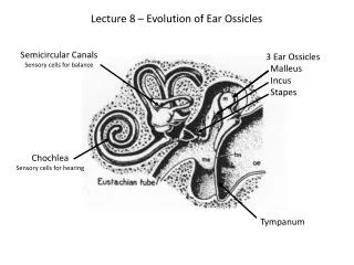
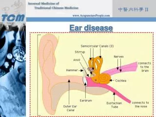
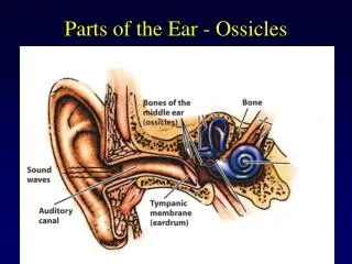
Ear ossicles. Malleus-Mallet(Hammer). Largest ossicle Parts Head Neck Anterior process Lateral process Handle. Parts. Head-body of incus Neck-pars flaccida, chorda tympani Anterior process- anterior ligament-petrotympanic fissure Lateral process-tympanic sulcus

E N D
Malleus-Mallet(Hammer) Largest ossicle Parts Head Neck Anterior process Lateral process Handle
Parts Head-body of incus Neck-pars flaccida, chorda tympani Anterior process- anterior ligament-petrotympanic fissure Lateral process-tympanic sulcus Handle-tympanic membrane tensor tympani muscle
Incus-anvil Body: head of malleus Long process: lentiform nodule- head of stapes Short process: fossa incudis-posterior wall of tympanic Cavity. Lentiform process/nodule: Head of Stapes - Incudo stapedial joint.
Stapes-stirrup Head- long process of Incus Neck- insertion of stapedius Anterior and posterior crus- Footplate- oval window/ fenestra vestibuli- annular ligament
Ossicular ligaments and Joints Malleus Anterior ligament Superior ligament Incus Posterior ligament Superior ligament Stapes Annular ligament Joints Incudomalleolar – saddle Incudostapedial joint-ball and socket
Otosclerosis Common hereditary disease Normal laminar bone is removed by osteoclasts and replaced by unorganised bone of greater thickness,vascularity and cellularity.
AUDITORYTUBE It is an osseo cartilagenous tube Communicates anterior wall of middle ear cavity with lateral wall of nasopharynx. Length: 36 mm It is directed downwards forwards and medially
AUDITORYTUBE Parts: Bony- 12 mm in length Cartilagenous - 24mm in length
Bonypart It has two ends Lateral end is broader which opens into anterior wall of middle ear. Medial end gives attachment to cartilagenous part of the auditory tube
Relations: Medially: Carotid canal which transmits ICA with sympathetic plexus Lateral: T.M.J, spine of sphenoid, chorda tympani nerve Superior: canal for tensor tympanic muscle
Cartilagenous part • Made up of triangular plate of fibrocartilage. • Situated in a groove on base of skull-sulcus tubae
Cartilaginous part • Apex of the cartilage is attached to medial end of bony part of tube.
Cartilaginous part • Base/Medial end of tube forms a projection of mucous membrane in lateral wall of nasopharynx -Tubal elevation.
Cartilaginous part • Base forms roof and medial wall of tube. • Rest of the area is filled by fibrous membrane.
Relations Anterolaterally: • Tensor Velipalatini • Mandibular nerve • Otic ganglion • Nerve to medial pterygoid • Middle meningeal artery • Chordatympani nerve
Posteromedially; • Apex of petrous temporal bone, LevatorVelipalatini muscle • Above Sulcustubae • Below Superior constrictor
Blood supply: a)Ascending pharyngeal artery b) Middle meningeal artery • Nerve supply: • Bony part; tympanic plexus • Cartilagenous part: Meningeal branch of mandibular nerve • Opening: Pharyngeal branch of pterygopalatine ganglion
Development: • Tubotympanic recess • Functions: • Equalises pressure outside and inside tympanic membrane • It is always open except and closed at rest and during deglutition • Muscles that open tube are • A) Salpingopharyngeus • B)Levatorvelipalatini • C) Tensor Velipalatini
ET Function Tests • VALSALVA TEST • Principle: positive pressure in the nasopharynx causes air to enter the Eustachian tube
In the immediate viscinity of the orifice of the eustachian tube (entrance middle ear), lies pharyngeal tonsil . Its obstruction, due to excessive tonsillar size (adenoids), may produce accumulation of fluid behind the tympanic membrane.