Download
1 / 29
320 likes | 1.02k Views
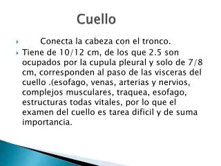
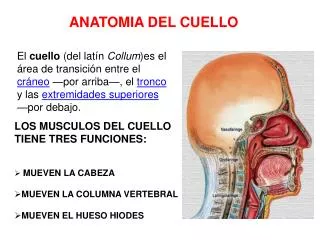
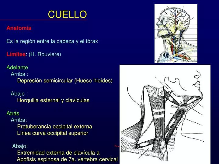
CUELLO. Anatomía Es la región entre la cabeza y el tórax Límites : (H. Rouviere) Adelante Arriba : Depresión semicircular (Hueso hioides) Abajo : Horquilla esternal y clavículas Atrás Arriba: Protuberancia occipital externa

E N D
CUELLO Anatomía Es la región entre la cabeza y el tórax Límites: (H. Rouviere) Adelante Arriba : Depresión semicircular (Hueso hioides) Abajo : Horquilla esternal y clavículas Atrás Arriba: Protuberancia occipital externa Línea curva occipital superior Abajo: Extremidad externa de clavícula a Apófisis espinosa de 7a. vértebra cervical
CUELLO Espacios del cuello La faringe puede dividirse en dos partes Superior o cefálico Inferior o cervical La división ocurre a nivel del borde inferior de la mandíbula o a nivel del hueso hioides
CUELLO Espacios del cuello Espacio Peri-faríngeo Un Retro-faríngeo Dos 2 Latero-faríngeo o maxilofaríngeo Diafragma estiloideo Pre-estiloideo Parotídeo Para-amigdalino Retro-estiloideo Carotídeo
CUELLO Espacios del cuello Por encima del borde inferior de la mandíbula Espacio Perifaríngeo o Parafaríngeo o Maxilo-faríngeo
CUELLO Espacios del cuello Por encima del borde inferior de la mandíbula Retrofaríngeo Laterofaringeo
CUELLO Espacios del cuello Por encima del borde inferior de la mandíbula Laterofaringeo Pre-estiloideo Retro-estiloideo
CUELLO Espacios del cuello Por encima del borde inferior de la mandíbula Laterofaringeo Pre-estiloideo
CUELLO Espacios del cuello Por encima del borde inferior de la mandíbula Laterofaringeo Pre-estiloideo -Para-amigdalino -Parotídeo Retro-estiloideo Carotídeo
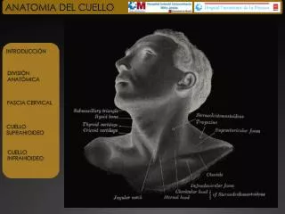
Espacios suprahioideos del cuello • e. Faríngeo mucoso • e. Parafaríngeo • e. Submandibular • e. Sublinguall • e. Cervical posterior • e. Carotídeo • e. Retrofaríngeo • e. Prevertebral • m. Geniohioideo y geniogloso • m. Hipogloso • m. Trapezio • m. Esternocleidomastideo • Proceso estiloides • e. Parotídeo • n. Facial • Glánd. Submaxilar • Gland. Parótida • Vena retromandibular • m. Milohioideo • Mandíbula Handbook of neck imaging, 1995
CUELLO • Fascias del cuello • Por debajo del borde inferior de la mandíbula • Piel • Tejido adiposo o celular subcutáneo • Fascia superficial • (envuelve el platisma) y • venas yugulares anteriores • Esternocleidomastoideo y Trapecio • Fascia o aponeurosis media • (envuelve músculos pre-laríngeos) • Fascia viseral que • (envuelve la tráquea • y el esófago y tiroides) • Fascia profunda • Pre-vertebral
CUELLO Arterias Carótida externa Tiroidea superior Faríngea ascendente Lingual Facial Occipital Auricular posterior Temporal superficial Maxilar
CUELLO Venas Seno lateral Vena yugular interna Vena yugular externa
CUELLO • Músculos del cuello • Laterales (5) • Superficiales • Platisma • Esternocleidomastoideo • Profundos • Escaleno anterior • Escaleno medio • Escaleno posterior
CUELLO • Músculos del cuello • Laterales (5) • Superficiales • Platisma • Esternocleidomastoideo • Profundos • Escaleno anterior • Escaleno medio • Escaleno posterior
RAM RAm CUELLO • Músculos del cuello • Anteriores (11) • Pre-vertebrales • Recto ant. mayor de la cabeza • Recto ant. menor de la cabeza • Largo del cuello • Infra-hioideos • Esterno-hioideo • Esterno-tiroideo • Omo-hioideo • Tiro-hioideo LC
CUELLO • Músculos del cuello • Anteriores (11) • Pre-vertebrales • Recto ant. mayor de la cabeza • Recto ant. menor de la cabeza • Largo del cuello • Infra-hioideos • Esterno-hioideo • Esterno-tiroideo • Omo-hioideo • Tiro-hioideo EH TH ET OH
CUELLO • Músculos del cuello • Anteriores (11) • Pre-vertebrales • Recto ant. mayor de la cabeza • Recto ant. menor de la cabeza • Largo del cuello • Supra-hioideos • Digástrico • Estilo-hioideo • Milo-hioideo • Genio-hioideo
CUELLO Triángulos del cuello Región parotídea Submentoniano posterior Submandibular Carotídeo Muscular Supra-clavicular
CUELLO Triángulos del cuello Región parotídea Submentoniano posterior Submandibular Carotídeo Muscular Supra-clavicular
CUELLO Niveles ganglionares Nivel I Nivel II Nivel V Nivel III Nivel IV Nivel VI
CUELLO Niveles ganglionares Nivel I Nivel V Nivel II Nivel III Nivel IV Nivel VI
Nivel I (Grupo submentonianos y submaxilar) hacia arriba: por el borde mandibular; hacia atrás: el vientre posterior del digástrico; hacia abajo: el hueso hioides; hacia delante: línea media
Nivel II (Grupo Yugular superior) De abajo hacia arriba desde el nivel del hueso hioides (referencia clínica) o bifurcación carotídea (referencia quirúrgica) hasta la base del cráneo. De adelante hacia atrás desde el vientre posterior del digástrico hasta el borde posterior del esternocleidomastoideo
Nivel III (Grupo Yugular Medio) Se extiende desde la escotadura cricotiroidea (referencia clínica) o músculo omohioideo (referencia quirúrgica) al borde inferior del nivel II.
Nivel IV (Grupo Yugular Inferior) Se extiende desde la clavícula al borde inferior del nivel III. Adelante desde el esternohioideo al borde posterior del esternocleidomastoideo.
Nivel V (Triángulo posterior) Limitado por el borde posterior del esternocleidomastoideo, el borde anterior del trapecio, y la clavícula.
Nivel VI (Compartimento Anterior) Desde los bordes internos o mediales de las láminas carótidas, El hueso hioides y la escotadura esternal