Download
1 / 74
750 likes | 885 Views
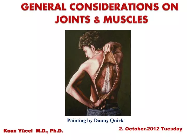
GENERAL CONSIDERATIONS ON JOINTS & MUSCLES. Painting by Danny Quirk. 2. October . 201 2 Tuesday. Kaan Yücel M.D., Ph.D . 1. GENERAL CONSIDERATIONS ON JOINTS. 1.1. CLASSIFICATION OF JOINTS 1.2. STABILITY OF JOINTS 1.3. JOINT VASCULATURE AND INNVERVATION.

E N D
GENERAL CONSIDERATIONS ON JOINTS & MUSCLES Painting by Danny Quirk 2. October.2012 Tuesday Kaan Yücel M.D., Ph.D.
1. GENERAL CONSIDERATIONS ON JOINTS 1.1. CLASSIFICATION OF JOINTS 1.2. STABILITY OF JOINTS 1.3. JOINT VASCULATURE AND INNVERVATION
2. GENERAL CONSIDERAITONS ON MUSCLES 2.1. Types of Muscles 2.2. Skeletal Muscles 2.2.1. Features of muscles 2.2.2. Muscle terminology 2.3. Contraction of muscles 2.4. Functions of muscles 2.5. FASCIA 2.6. Nerves and arteries of muscles
Arthrology Greek a rqron joint –logy • science concerned with the • anatomy, function, dysfunction and treatment of joints.
according to the tissues that lie between the bones: Fibrous joints Cartilaginous joints Synovial joints Classification of Joints
Fibrous joints • Bones are united by fibrous tissue. • Suturesof the cranium
Fibrous joints • Syndesmosistype of fibrous joint • unites the bones with a sheet of fibrous tissue • either a ligament or a fibrous membrane • partially movable • The interosseous membrane in the forearm is a sheet of fibrous tissue that joins the radius and ulna in a syndesmosis.
Fibrous joints • Syndesmosistype of fibrous joint • unites the bones with a sheet of fibrous tissue • either a ligament or a fibrous membrane • partially movable • The interosseous membrane in the forearm is a sheet of fibrous tissue that joins the radius and ulna in a syndesmosis.
Cartilaginous joints Bones are united by hyaline cartilage or fibrocartilage.
Cartilaginous joints Pimarycartilaginous joints-synchondroses hyaline cartilage- growth of a bone duringearly life Secondary cartilaginous joints-symphyses strong, slightly movable joints united by fibrocartilage
Synovial joints • Most common type of joints • Bones united by a joint capsule enclosing an articular cavity. • Providefree movement between the bones they join. • Joint cavity • potential space • contains lubricating synovial fluid, secreted by the synovial membrane. • Articular cartilage • articular surfaces are covered by hyaline cartilage • Articular capsule • surrounds the joint and formed of two layers.
Articular capsule: • surrounds the joint • two layers. • Fibrous capsule • Synovial membrane Some synovial joints have other distinguishing features, such as a fibrocartilaginousarticular disc or meniscus, which are present when the articulating surfaces of the bones are incongruous.
Ligaments • a cordorband of connectivetissueunitingtwostructures. • Articularcapsulesareusuallystrengthenedbyarticularligaments. • Connect thearticulatingbonestoeachother. • limit theundesiredand/orexcessivemovements of thejoints.
Articulardisc: Help toholdthebonestogether. Labrum: A fibrocartilaginous ring whichdeepensthearticularsurfaceforone of thebones.
Bursa • Flattenedsacsthatcontainsynovialfluidtoreducefriction. • Wallsare separated by a film of viscous fluid. • Foundwherever tendons rub against bones, ligaments, or other tendons.
Stability of Joints Negativepressure within the joint cavity Shape, size, and arrangement of the articular surfaces Ligaments Tone of the muscles around the joint
Jointvasculatureandinnvervation • Joints receive blood from articular arteries that arise from the vessels around the joint. • Articular veins are communicating veins that accompany arteries (L. venae comitantes) and, like the arteries, are located in the joint capsule, mostly in the synovial membrane. • Joints have a rich nerve supply provided by articular nerves with sensory nerve endings in the joint capsule.
Types of synovial joints • according to shape of articulating surfaces- type of movement they permit • Plane joints • uniaxialjoints- glidingorsliding • acromioclavicular joint • 2. Hinge joints • uniaxialjoints- flexion & extension • knee & elbowjoints
Types of synovial joints 3. Saddle joints biaxialjoints- flexion & extension, abduction & adduction carpometacarpal joint at the base of the 1st digit (thumb) 4. Condyloid (ellipsoid type) biaxialjoints- flexion & extension, abduction & adduction metacarpophalangeal joints (knuckle joints) radiocarpal joint (wrist)
Types of synovial joints 5. Ball and socket joints (spheroidaljoints) multiple axes and planes: flexion and extension, abduction and adduction, medial and lateral rotation, and circumduction hip & shoulderjoints
Types of synovial joints 6. Pivot joints uniaxialjoints- rotationaround a centralaxis proximal& distal radioulnar joints
TendonSheath • A layerof thesynovialmembranearound a tendon. • Permitsthetendontomove.
EXAMINATION OF JOINTS The clinician should assess the normal range of movement of all joints. When the bones of a joint are no longer in their normal anatomic relationship with one another, then the joint is said to be dislocated. . Examination of theshoulderjoint
Dislocation of Joints Some joints are particularly susceptible to dislocation because of: lack of support by ligaments the poor shape of the articular surfaces, the absence of adequate muscular support. The shoulder joint, temporomandibular joint, &acromioclavicularjoints .
Damage to Ligaments • Ligaments prone to excessive stretching &even tearing and rupture. • If possible, the apposing damaged surfaces of the ligament are brought together by positioning and immobilizing the joint. • In severe injuries, surgical approximation of the cut ends may be required. .
Osteoarthritis Synovial joints are well designed to withstand wear, but heavy use over several years can cause degenerative changes. Some destruction is inevitable during such activities as jogging, which wears away the articular cartilages and sometimes erodes the underlying articulating surfaces of the bones. .
Osteoarthritis The normal aging of articular cartilage begins early in adult life and progresses slowly thereafter, occurring on the ends of the articulating bones, particularly those of the hip, knee, vertebral column, and hands. .
Osteoarthritis • Degenerative joint disease or osteoarthritisis often accompanied by stiffness, discomfort, and pain. • Osteoarthritis is common in older people and usually affects joints that support the weight of their bodies (e.g., the hips and knees). .
Arthroscopy Cavityof a synovial joint can be examined by inserting a cannula and an arthroscope (a small telescope) into it. Enables to examine joints for abnormalities, such as torn menisci (partial articular discs of the knee joint). Some surgical procedures can also be performed. .
interested in allthemuscles in the body • Musculus (muscle) mus-mouse; musculus- little mouse. • So called because the shape and movement of some muscles (notably biceps) were thought to resemble mice. • If you bend and straighten your arm at the elbow, you should see the front of the upper arm move under the skin. To the ancient Romans this movement resembled a little mouse scurrying beneath the skin. myology
Types of Muscles • based on distinct characteristics • Functional • voluntary vs. involuntary • Histological • striated vs. smooth or unstriated • Anatomical (location) • @ body wall (soma) and limbs • @ hollow organs (viscera) or blood vessels
Skeletal striated muscle voluntarysomatic muscle gross skeletal muscles that compose the muscular system moving or stabilizing bones and other structures (e.g., the eyeballs). Innervatedby the somatic nervous system.
Cardiac striated muscle involuntary visceral muscle forms the walls of the heart and adjacent parts of the great vessels. pumps blood.
Smooth muscle (unstriated muscle) involuntary visceral muscle forms part of the walls of most vessels and hollow organs (viscera) moving substances through them coordinated sequential contractions (pulsations or peristaltic contractions). Innervatedby the autonomic nervous system.
FEATURES OF SKELETAL MUSCLES HEAD OR BELLY fleshy, reddish, contractile portions TENDONwhite non-contractile portions composed mainly of organized collagen bundles, that provide a means of attachment.
Mostskeletalmusclesattachto Directlyorindirectlytobones Cartilages Ligaments Fascias orcombinations of theonesabove Sometoorgans (eyeball)/skin (facialmuscles)/mucousmembranes(intrinsictonguemuscles)
Muscles are organs of locomotion (movement) also: provide static support give form to the body provide heat
Sometendons form flatsheetsaponeuroses anchor the muscle to the skeleton to deep fascia toaponeurosisof another muscle
Many terms provide information about a structure's Shape Size Location Function Resemblanceof one structure to another
Basisof function Bones attachedto Abductordigitiminimimuscleabducts the little finger. Sternocleidomastoidmuscle (G. kleidos, bolt or bar, clavicle) attaches inferiorly to the sternum and clavicle and superiorly to the mastoid process of the temporal bone of the cranium. Levatorscapulae elevates the scapula (L. shoulder blade).
Descriptive names Deltoid muscle triangular, like the symbol for delta, the fourth letter of the Greek alphabet. -oid“like”; deltoid means like delta.
Position • medial, lateral, anterior, posterior • Length • brevis, short; longus, long • Shape • piriformismuscle • pear shaped (L. pirum, pear + L. forma, shape or form).
Location • temporalismuscle • in the temporal region (temple) of the cranium (skull).
CLASSIFICATION OF MUSCLES accordingtotheirshapes Flat muscles parallel fibers often with an aponeurosis External obliquemuscle broad flat muscle Sartorius narrow flat muscle with parallel fibers longestmuscle in the body
feather-like (L. pennatus, feather), arrangement of fasicles Unipennate Extensor digitorum longus Bipennate Rectus femoris Pennate muscles Multi-pennate Deltoid
spindle shaped with a round, thick belly (or bellies) and tapered ends Fusiformmuscles