Download
1 / 44
440 likes | 543 Views
Acute Injury to the Spine and Brain. Dr.Fahad Al Bader. How to read a spinal x-ray. ABCS 1-Alignment 2-Bone integrity 3-Cartilage spaces (disc spaces, atlanto -axial space) 4-Soft tissues ( prevetebral space 30% C3 100% C7). Spinal x-ray abnormalities.

E N D
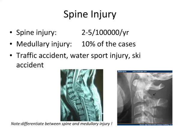
Acute Injury to the Spine and Brain Dr.Fahad Al Bader
How to read a spinal x-ray ABCS 1-Alignment 2-Bone integrity 3-Cartilage spaces (disc spaces, atlanto-axial space) 4-Soft tissues (prevetebral space 30% C3 100% C7)
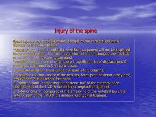
Spinal x-ray abnormalities When reading a spinal x ray look for: Alignment and Sulbluxation Paravertebral swelling Collapsed vertebra Spondylolysis Spondylolisthesis Retrolisthesis
Scottie Dog Neck : pars interarticularis eye : pedicle hindlegs : spinous process nose : transverse process ear : superior articular facet forelegs : inferior articular facet
How to read a Brain CT scan 1-Site of the lesion : Intra or extra axial 2- ventricles 3-skull and orbit
Sites of Normal Calcification in the Brain: 1-pineal gland 2-choroid plexus 3-basal ganglia
Hyperdense Lesions Acute Hematoma Look for fractures Is the patient hypertensive?
Epidural VS Subdural Hematoma Epidural Subdural • Young Men (20-40's) – Head Trauma frequent – Also, dura (periosteum) more adherent in older people • Acute presentation • Skull fracture (90%) • Bi-convex, hyperdense- limited by sutures. • Arterial (meningeal vessels) • Acute to Chronic • Older age group • Concave layer • Fracture +/- • Cross sutures • Venous (bridging veins)
Brain Herniation Subfalcine : theinnermost part of the frontal lobe is scraped under part of the falxcerebri. Cingulateherniation can be caused when one hemisphere swells and pushes the cingulategyrus by the falxcerebri Uncal: the innermost part of the temporal lobe, the uncus, can be squeezed so much that it goes by the tentorium and puts pressure on the brainstem, most notably the midbrain
Types of Brain Edema Vasogenic: Due to a breakdown of tight endothelial junctions which make up the blood-brain barrier (BBB) - hydrostatic (malignant HTN) - brain cancer (VEGFdexamethasone) - high altitude Cytotoxic : - the BBB remains intact - it is due to the derangement in the celllar metabolism - inadequate functioning of the Na and K pump -various intoxications (dinitrophenol, triethyltin, hexachlorophene, isoniazid) -Reye's syndrome, -severe hypothermia, -early ischemia, -encephalopathy, early stroke or hypoxia, cardiac arrest, pseudotumorcerebri, and cerebral toxins.