Download
1 / 17
170 likes | 298 Views
1. A chemical process where there is a net gain of electrons is called _______________. A chemical process where there is a net loss of electrons is called _________________. 2. Enzymes are catalysts, they often _____________ the free energy of a reaction by favoring a transition state.

E N D
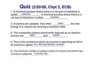
1. A chemical process where there is a net gain of electrons is called _______________. A chemical process where there is a net loss of electrons is called _________________. 2. Enzymes are catalysts, they often _____________ the free energy of a reaction by favoring a transition state. 3. This nucleotide cofactor prominently featured as an electron carriers are _____________ and ______________. 4. This is the constant at which an enzyme is operating at half of its maximum speed. _____________________ 5. The maximum number of catalytic cycles an enzyme can perform per unit time is called the _____________. Quiz (2/20/08, Chpt 3, ECB) reduction oxidation lower NAD+ NADP+ Km, Michalis-Menten constant Turnover rate
FIONA Fluorescence Imaging with One Nanometer Accuracy (1.5 nm, 1-500 msec)
Fluorescence l out 8 nm Techniques needed Specificity to look at heads Nanometer spatial localization Second temporal resolution Single Molecule sensitivity Single Molecule Photostability l in How to get nanometer localization with visible photons?
Imaging Single Moleculeswith very good S/N Total Internal Reflection Microscopy TIR- (q > qc) Exponential decay dp=(l/4p)[n12sin2i) - n22]-1/2 For water (n=1.33) to air (n=1.0): what is TIR angle? For glass (n=1.5), water (n=1.33): what is TIR angle? what is penetration depth? >57° dp = 58 nm With dp = 58 nm , can excite sample and not much background.
Laser Sample Objective Sample Dichroic Laser Objective Filter Filter Lens Lens CCD Detector CCD Detector Wide-field Objective-TIR Wide-field, Prism-type, TIR Microscope Experimental Set-up for TIR(2 set-ups) No! In one case, sample is “upside-down.” Does this make a difference?
center width Diffraction limited spot Width of l/2 ≈250 nm Accuracy of Center = width/ S-N = 250 nm / √104= 2.5 nm= ± 1.25nm Enough photons (signal to noise)…Center determined to ~1.3 nm Dye last 5-10x longer -- typically ~30 sec- 1 min. (up to 4 min)
center width = derived by Thompson et al. (Biophys. J.). How well can you localize?What does it depend on?(3 things) 1. # of Photons Detected (N) 2. Pixel size of Detector(a) 3. Noise (Background) of Detector (b) (includes background fluorescence and detector noise)
DNA Sample 18mer Biotinylated DNA Cy-3 Dye 3’ Streptavidin Biotinylated BSA coverslip coverslip Move stage In 8 nm, 16 nm, 37 nm increments Experimental Setup: Imaging Single MoleculesCy3-DNA Immobilized on coverslip
m = 28.52nm 10 800 s = 1.24nm m = 28.55nm Ex. no. of steps 5 600 0 26 28 30 32 poissonian step size (nm) m = 29.08nm s = 1.32nm 400 m = 28.55nm Ex. 10 200 no. of steps 5 0 26 28 30 32 regular step size (nm) 0 0 20 40 60 80 100 Time (sec) Data: Model System: 30 nm Artificial Steps Position (nm)
12 nm steps m = 12.47nm s = 1.37nm m = 12.35nm Ex. 5 150 4 3 no. of steps 2 120 1 0 10 11 12 13 14 15 regular step size (nm) m = 12.26nm 90 s = 1.39nm m = 12.35nm Ex. 60 3 2 no. of steps 30 1 0 10 11 12 13 14 poissonian step size (nm) 0 0 10 20 30 40 50 Time (sec) Position (nm)
8 nm steps m = 7.63nm s = 0.93nm 5 m = 7.45nm Ex. 4 100 3 2 1 0 80 6 7 8 9 60 m = 6.42nm Position (nm) s = 1.09nm m = 6.52nm Ex. 40 5 4 3 no. of steps 20 2 1 0 4 5 6 7 8 9 regular step size (nm) 0 0 10 20 30 40 50 Time (sec) no. of steps poissonian step size (nm)
Myosin V Labeling on Light Chain: Expected Step Sizes 37 nm 37/2 nm x 37-2x 37+2x Center of mass 0 nm 74 nm Expected step size Hand-over-hand: Head = 2 x 37 nm= 74, 0, 74 nm CaM-Dye: 37-2x, 37+2x, … Inchworm: always Scm = 37 nm
A Single Myosin V moving [ATP] = 300 nM (Low) 86 nm pixel 37 nm or 74 nm?
Myosin V steps: 74 nm +/- 5nm [ATP] = 300 nM higher [ATP] (>400 nM) +/- 1.3 nm +/- 1.5 nm +/- 1.3 nm
74-0 nm Steps: Detecting 0 nm Intermediate by Kinetics B A k1 k2 A u sec (t-u) sec 74-x nm x nm 74 nm (t sec) observable! 40-30 nm and 50-20 nm dwell time histogram k = 0.302 R2 = 0.979 Nmol=12 Nstep=191 74-0 nm dwell time histogram k1 =0.3281 Nmol=37 k2 = 0.3278 Nstep=361 R2 = 0.983 k1=k2 k = 0.3279 ± 0.007 A [ATP] = 300 nM
Class evaluation • What was the most interesting thing you learned in class today? • 2. What are you confused about? • 3. Related to today’s subject, what would you like to know more about? • 4. Any helpful comments. Answer, and turn in at the end of class.