Download
1 / 53
570 likes | 1.14k Views
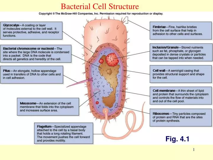
Bacterial Cell Structure. Fig. 4.1. Cytoplasm. dense gelatinous solution of sugars, amino acids, & salts 70-80% water serves as solvent for materials used in all cell functions. Chromosome.

E N D
Bacterial Cell Structure Fig. 4.1
Cytoplasm • dense gelatinous solution of sugars, amino acids, & salts • 70-80% water • serves as solvent for materials used in all cell functions
Chromosome • single, circular, double-stranded DNA molecule that contains all the genetic information required by a cell • DNA is tightly coiled around a protein, aggregated in a dense area called the nucleoid
plasmids • small circular, double-stranded DNA • free or integrated into the chromosome • duplicated and passed on to offspring • not essential to bacterial growth & metabolism • may encode antibiotic resistance, tolerance to toxic metals, enzymes & toxins • used in genetic engineering- readily manipulated & transferred from cell to cell
Ribosomes • made of 60% ribosomal RNA & 40% protein • consist of 2 subunits: large & small • procaryotic differ from eucaryotic ribosomes in size & number of proteins • site of protein synthesis • All cells have ribosomes.
Inclusions, granules • intracellular storage bodies • vary in size, number & content • bacterial cell can use them when environmental sources are depleted • Examples: glycogen, poly-b-hydroxybutyrate, gas vesicles for floating, sulfur and polyphosphate granules
Cytoplasmic membrane Protoplast Spheroplast L forms
4 groups based on cell wall composition • Gram positive cells • Gram negative cells • Bacteria without cell walls • Bacteria with chemically unique cell walls
Gram positive Gram negative
Gram positive Gram negative Fig 4.16
Lipopolysaccharide O-antigen Highly variable n • Core • Heptoses • Ketodeoxyoctonic acid • Lipid A • Glucosamine disaccharide • Beta hydroxy fatty acids (Hydroxy myritic Acid)
Lipoteichoic acid Peptidoglycan-teichoic acid Cytoplasmic membrane Cytoplasm Lipopolysaccharide Porin Outer Membrane lipoprotein Periplasmic space Inner (cytoplasmic) membrane Cytoplasm
r r r r r r r r r r Gram Positive Cell Envelope Lipoteichoic acid Peptidoglycan-teichoic acid Cytoplasmic membrane Cytoplasm
Glycocalyx • Coating of molecules external to the cell wall, made of sugars and/or proteins • 2 types • capsule - highly organized, tightly attached • slime layer - loosely organized and attached • Functions • attachment • inhibits killing by white blood cells • Receptor (K antigen)
Monotrichous lophotrichous amphitrichous peritrichous
Fimbrae • Adhesion to other cells and surfaces
pili • rigid tubular structure made of pilin protein • found only in Gram negative cells • Functions • joins bacterial cells for DNA transfer (conjugation) • adhesion
Major Taxonomic Groups of Bacteria • Gracilicutes – gram-negative cell walls, thin-skinned • Firmicutes – gram-positive cell walls, thick skinned • Tenericutes – lack a cell wall & are soft • Mendosicutes – archaea, primitive procaryotes with unusual cell walls & nutritional habits
Grwth in Bacteria • Temperature • Nutrients • pH • Osmotic pressure
Temperature • Minimum temperature – lowest temperature that permits a microbe’s growth and metabolism • Maximum temperature – highest temperature that permits a microbe’s growth and metabolism • Optimum temperature – promotes the fastest rate of growth and metabolism
Bacterial Metabolism • Phototroph • Photoautotroph (Photolitotroph) • Photoheterotroph (Photoorganotroph) • Chemotroph • Chemoautotroph (Chemolitotroph • Chemoheterotroph (Chemoorganotroph)
Stages of metabolism in chemoheterotrophic bacteria • Digestion • Absorption(Passive and active transportation) • Preparation for oxidation • Oxidation
Fermentation • Incomplete oxidation of glucose or other carbohydrates in the absence of oxygen • Uses organic compounds as terminal electron acceptors • Yields a small amount of ATP • Production of ethyl alcohol by yeasts acting on glucose • Formation of acid, gas & other products by the action of various bacteria on pyruvic acid
Continuous Culture, Chemostat Chemostats are a means of keeping a culture in log phase indefinitely.
Methods in bacterial identification • Microscopic morphology • Macroscopic morphology – colony appearance • Physiological / biochemical characteristics • Chemical analysis • Serological analysis • Genetic & molecular analysis • G + C base composition • DNA analysis using genetic probes • Nucleic acid sequencing & rRNA analysis
Bacterial Colonies • Standard Bacterial Count • Colony-Forming Units • Plaque-Forming Units • Spread Plate • Pour Plate • Soft-Agar Overlay