Download
1 / 23
230 likes | 332 Views
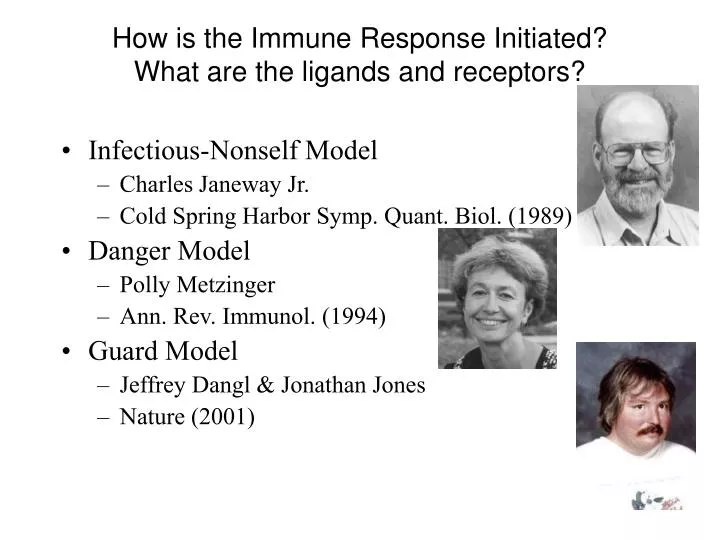
How is the Immune Response Initiated? What are the ligands and receptors?. Infectious-Nonself Model Charles Janeway Jr. Cold Spring Harbor Symp. Quant. Biol. (1989) Danger Model Polly Metzinger Ann. Rev. Immunol. (1994) Guard Model Jeffrey Dangl & Jonathan Jones Nature (2001).

E N D
How is the Immune Response Initiated?What are the ligands and receptors? • Infectious-Nonself Model • Charles Janeway Jr. • Cold Spring Harbor Symp. Quant. Biol. (1989) • Danger Model • Polly Metzinger • Ann. Rev. Immunol. (1994) • Guard Model • Jeffrey Dangl & Jonathan Jones • Nature (2001)
Infectious-Nonself Model • Detection of conserved molecular patterns (pathogen-associated molecular patterns, PAMPs) by pattern recognition receptors (PRR). • Upon recognition of PAMPs by … • Membrane-associated PRR: signaling pathways that induce antimicrobial effectors and inflammation is activated. • Soluble PRRs: pathogens bound and flagged for destruction by phagocytosis or the complement system.
Identification of PRR in Drosophila • Drosophila produces 7 structurally diverse, cationic and predominantly membrane active AMPs. • Activity Spectra: • Fungi: Drosomycins, Metchnikowin • Gram-positive bacteria: Defensin • Gram-negative bacteria: Attacins, Cecropins, Drosocin, Diptericins • The promoter regions of these AMP genes contain nucleotide motifs similar to mammalian binding sites for NF-kB/Rel proteins. • The Drosophila NF-kB/Rel ortholog is Dorsal, had been genetically identified as a regulator of dorsoventral patterning in the early embryo. • In 1996, Hoffmann’s group reported that loss-of-function mutations in the Toll receptor compromised the survival of flies faced with fungal infection and the challenge-dependent transcription of the antifungal peptide Drosomycin.
Identification of PRR in mammals • In 1994, the first mammalian TLR, TLR1 was cloned and was identified as a homolog of the Drosophila Toll [DNA Res. 1, 27]. It was designated as TIL (Toll/IL-1 receptor-like) by Taguchi et al in 1996 [Genomics 32, 486] and was suspected to have a developmental function as the immune function of Toll had not been demonstrated. • In 1997, Janeway and Medzhitov successful cloned a human homologue of Drosophila Toll and showed this Toll-like receptor (TLR) activate NF-kB [Nature 388, 394]. • The immune relevance of TLR was shown in 1998 when Bruce Buetler and colleagues showed that defective LPS signaling in C3H/HeJ and C57BL/10ScCr mice was a consequence to mutation in Tlr4 [Science 282, 2085]. • Akira’s group, by means of gene targeting, determined the main microbial specificity of TLR2, TLR6 and TLR9.
Human Toll-like Receptors Members of the interleukin-1 receptors (IL-1Rs) family. They share a conserved cytoplasmic region known as the Toll/IL-1R (TIR) domain. But the extracellular portion of the TLRs contains a leucine-rich repeat (LRR) motif whereas that of the IL-1Rs contains three immunoglobulin domains.LRR domains may be directly involved in the recognition of PAMPs.TLR7 & TLR9 are in intracellular compartments.Individual TLR can interact with several structurally unrelated ligands of exogenous and endogenous origin.
TLR Signal Transduction pathway All TLR signal through MyD88.Individual TLR also induce pathogen-specific immune response.TLR2 & TLR4 also signal through TIRAP/Mal.TLR3 (?TLR4) also signal through TRIF.Induction of IFNa/b expression through IRF3 by TLR3 & TLR4 is MyD88-ndependent.
What features distinguish Gram +ve and Gram -ve Peptidoglycan (PGN)? Gram-positive Gram-negative
The Danger Model • The immune system is activated in response to substances that cause damage, rather than those that are simply foreign. These alarm/danger signals are molecules or molecular structures, released or produced by cells undergoing stress or abnormal death. These signals are perceived by resting APCs. • The alarm signals can be either endogenous or exogenous, intracellular or extracellular/secreted. They may function as primal initiators or simply give positive-feedback to enhance or modify an ongoing response.
Empirical Support for the Danger Model • Resting DCs are activated by cells killed by acute necrotic death but no by cells dying of physiological apoptotic death [Nat. Med. 5, 1249 (99); J. Exp. Med. 191,423 (00)]. Signals produced by necrotic cells are heat shock proteins gp96 and hsp70 [Int. Immunol. 12, 1539 (00)]. • Crystalline, but not soluble, uric acid can activate DCs [Nature 425, 516 (03)].
Gene-for-Gene Hypothesis by H.H. Flor Disease resistance in plants requires two complementary genes: an avirulence (Avr) gene in the pathogen and a matching, resistance (R) gene in the host.Suggests a receptor-ligand model in which R-protein-mediated recognition of the pathogen-derived Avr products leads to hypersensitive response (HR) in plant. This hypothesis has led to the identification of many R-Avr protein pairs from plants and pathogens. However, with the exception of the in vitro interaction between Pi-ta (a rice R protein) and AvrPita from the fungal pathogen Magnaporthe grisea, no direct interactions between Avr and R protein has been demonstrated.
Identification of R genes in Plants • Prior to results obtained in animals, in 1993, Greg Martin cloned the Pto gene in tomato confers resistance to races of Pseudomonas syringae pv. tomato that carry the avirulence gene avrPto. • In 1994, the Ausubel and Staskawicz groups clones the resistance gene RPS2 from Arabidopsis [Science 265, 1856 & Cell 78, 1089]. In the same year, Barbara Baker’s group cloned the N gene from tobacco [Cell, 78, 1101]. • The N and RPS2 proteins, like Toll and TLRs has the LRR and TIR domains. They define a large group of cytoplasmic R proteins called the NBS-LRRs. They contain a central nucleotide binding site and a C-terminal LRRs called the NBS-LRR. • A third class of R genes are membrane-anchored glycoprotein with extracytoplasmic LRRs.
Recognition of AvrPto by tomato NB-LRR protein (Prf) is mediated by Pto
The Guard Model R proteins associate physically and specifically with cellular targets (or “guardee”) of bacterial type III effectors (Avr). The interaction between guardee and Avr is recognized by the R protein, which is thus activated to initiate disease resistance. Guardees are likely to be plant defense components or host proteins whose function is modified to nourish the extracellular bacterial pathogen. In the absence of a specific R protein, the host target is not guarded from the virulence function of Avr, and disease ensues.
Evidence Supporting the Guard HypothesisCell 108, 743 (02); 112, 369 (03); 112, 379 (03) AvrRpt2 is a Cysteine Protease [Mol. Micro. 49,1537 (03)]