Download
1 / 42
420 likes | 620 Views
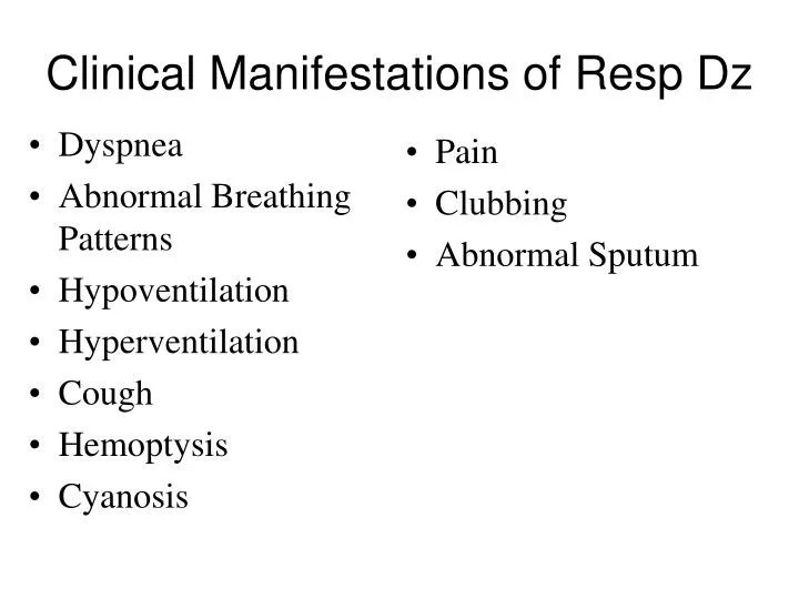
Pain Clubbing Abnormal Sputum. Clinical Manifestations of Resp Dz. Dyspnea Abnormal Breathing Patterns Hypoventilation Hyperventilation Cough Hemoptysis Cyanosis. Dyspnea. Difficulty breathing, Can’t catch my breath, Short of breath, air hungry No exact cause

E N D
Pain Clubbing Abnormal Sputum Clinical Manifestations of Resp Dz • Dyspnea • Abnormal Breathing Patterns • Hypoventilation • Hyperventilation • Cough • Hemoptysis • Cyanosis
Dyspnea • Difficulty breathing, Can’t catch my breath, Short of breath, air hungry • No exact cause • Length/tension inappropriateness theory • Chemoreceptors • Lung receptors • S/S: flaring nostrils, accessory muscle use, retractions, • DOE: early sign • Orthopnea, PND
Abnormal Breathing Patterns • Normal • tidal volume 400 – 800 ml; 6 – 20/min • Short expiratory pause • Sigh breaths 10-12 per hour • Kussmaul (strenuous exercise or acidosis) • Large tidal volume; rapid rate • Labored (obstructed) breathing • Slow rate, large tidal volume, prolonged insp or exp • Restricted breathing • Rapid rate, small tidal volume
Abnormal Breathing Patterns • Panting: exercise (small Kussmaul) • Gasping: shock, cerebral hypoxia • Sighing: anxiety • Cheyne-Stokes: slowing of blood to brain stem • Alternating deep and shallow followed by apnea
Hypoventilation Hyperventilation • Minute volume: (tidal volume) x (resp rate) • Hypoventilation: in relation to metabolic demands • Hypercapnia (PaCO2 > 45) Resp Acidosis • Easy to overlook • Causes: Somnolence, disorientation, Secondary hypoxemia • Hyperventilation • Hypocapnia (PaCO2 < 35) Resp Alkalosis • Caused by: Anxiety, head injury, inadequate oxygenation
Cough • Physiologic reflex: remove mucous and foreign particles from airway • Most initiated in Larnyx and bronchotracheal tree • Mechanical or chemical irritants • Others initiated: stomach, EAC, pericardium, pleura, • Acute: resolves 2-3 weeks • URI, allergic rhinitis, acute bronchitis, pneumonia, HF, Pulmonary embolus, aspiration • Chronic: last >3 weeks (or 7-8 weeks) • Postnasal, asthma, GERD, smokes, cancer, ACEI
Hemoptysis • Coughing up bloody secretions • Usually bright red or pink, alkaline pH • Frothy sputum • Etiology • Bronchiectasis, lung cancer, bronchitis, pneumonia • Worldwide: Tuberculosis • Amount and onset
Cyanosis • Desaturation of 5 g/dl of hemoglobin • Causes • Decreased arterial oxygenation • Right to left shunts • Decreased CO • Cold environments • Anxiety • Adults: not evident until extreme hypoxemia • Lips, buccal mucosa, nail beds
Pain • Pleural (Pleuritic) Pain • Caused by movement of inflamed pleura • Sharp, well localized • Friction rub • Also present in infarction d/t Pulmonary embolism • Pulmonary pain: central chest • Esp after coughing • Pulmonary hypertension • Pain chest wall: rib, muscle, or cartilage • Can mimic pleural pain
Clubbing • Selective bulbous enlargement of distal segment of a digit • Graded 1 – 5 on severity • Associated with diseases that impair oxygenation • Cystic fibrosis • Pulmonary fibrosis • Lung abcess • Congenital heart disease • Lung cancer
Abnormal Sputum • Color • Consistency • Amount • Odor • Sputum is not the same as saliva!!!!
Pleural Abnormalities Abcess & Cavitation Pulmonary fibrosis Chest Wall restriction Flail chest Inhalation D/Os Systemic D/Os Conditions Caused by Resp Disease or Injury • Hypercapnia • Hypoxemia • Acute Respiratory Failure • Pulmonary Edema • Aspiration • Atelectasis • Bronchiectasis • Bronchiolitis
Hypercapnia • PaCO2 > 45 • Etiology: Decreased drive to breathe or inability to ventilate in response to drive • 1. Depression of resp center by drugs • 2. Diseases or injury to medulla • 3. Abnormalities in spinal conduction (poliomyelitis) • 4. Diseases of neuromuscular junction (myasthenia gravis or muscular dystrophy) • 5. Thoracic abnormalities • 6. Large airway obstructions • 7. Increased work of breathing
Hypercapnia • Clinical manifestations: • Primarily through decreased pH → resp acidosis • Electrolyte imbalance (K+ esp) • Arrhthymias • Cerebral artery dilation → intracranial pressure • Somnolence • Coma • Hypoxemia • Loss of primary drive to breathe
Hypoxemia • Reduced oxygenation of blood (not tissue) • Etiology: • 1. Decreased O2 content of inspired air • Usually high elevation • Administer oxygen (or polycythemia) • 2. Hypoventilation • Often 2º hypercapnia • 3. Diffusion abnormalities • Thickened alveolar membrane or decreased surface area • Edema, Fibrosis, Emphysema • Usually not associated with hypercapnia
Hypoxemia • Etiology Cont • 4. Abnormal V/Q ratios • Most common cause • Shunting: asthma, pulmonary edema, pneumonia • Pulmonary right-to-left shunt: blood is not oxygenated d/t shunting; does not respond to ↑ oxygen • ARDS, respiratory distress of the newborn • Dead space: pulmonary embolism
Hypoxemia • Patho/Manifestations • Usually assoc w/ hyperventilation and resp alkalosis • Widespread tissue injury • Hypoxemic pulmonary vasoconstriction • Acute s/s: cyanosis, confusion, edema, ↓UOP
Acute Respiratory Failure • Inadequate gas exchange • PaO2 ≤ 50 mmHg OR • PaCO2 ≥ 50 mmHg with pH ≤ 7.25 • Etiology: injury to lungs, airway, chest wall, or indirect damage (brain or nerves) • If ARF is primarily hypercapnic, then ventilatory assistance is needed (bag or vent) • If ARF is primarily hypoxemi, then O2 needed • If mixed, then need both
Pulmonary Edema • Usually lung is fairly dry • Balance of hydrostatic and oncotic pressure • Lymphatic drainage • Surfactant repels water from alveoli • Most common etiology is cardiac • Left sided failure results in backup of fluid • Lymphatic drainage can manage small amount • Eventually, lymphatic drainage becomes saturated, and edema develop • Usually occurs when left atrial pressure ≥ 20 mmHg • Can occur at lower temps if ↓ oncotic pressure
Pulmonary Edema • Other Etiology • Increased pulmonary permeability (usually capillary injury or inflammation) • ARDS, inhalation toxic gas (ammonia) • Decreased lymphatic drainage • Cancer, fibrotic tissue, increased venous pressure of large pulmonary veins (heart failure)
Aspiration • Passage of solid or liquid particles into lung • Usually occurs with impaired swallowing, cough, or level of consciousness • Substance abuse, sedation, anesthesia, seizure, stroke, Myasthenia Gravis, Guillain-Barre • Enteral feeding • Tracheoesophageal fistula
Aspiration • Manifestations: depend on what was aspirated • Large chunks of food may completely occlude a bronchus (or trachea in small children) • Low pH fluids/food may cause local inflammation that leads to bronchiectasis (surgery required)
Aspiration • Aspiration pneumonia (especially if oral cavity colonized with bacteria) • Pneumonitis • Bronchial damage (loss of cilia action, bronchospam, inflammation) • Alveolar hemorrhage, fibrosis, atelectasis • Clinical manifestations • Choking, cough, vomiting, fever, dyspnea, wheezing • Recurrent lung infections, chronic cough
Aspiration • Prevention is better than treatment • NURSING CARE!!!!!! • Raise HOB, don't eat reclining, thickened fluids, set enteral feedings slower, check NG tube placement • Treatment • Aspiration pneumonitis has 50% mortality rate • Bronchoscopy • NG suction • Mechanical ventilation, O2, PEEP • Antibiotics is indicated
Atelectasis • Collapse of lung tissue • Compression atelectasis • External pressure: tumor, pleural effusion, abdominal distension • Absorption atelectasis • Gradual collapse 2° hypoventilation or obstructed airway • Manifestations • Dyspnea, cough, fever, leucocytosis
Atelectasis • Treatment • Compression: relieve compression • Absorption: deep breathing • Promotes ciliary clearing of secretions • Stabilizes alveoli by spreading surfactant • Permits collateral ventilation through pores of Kohn
Bronchiectasis • Persistent abnormal dilation of bronchi • Usually occurs w/ other respiratory conditions • Also occurs with systemic disorders • AIDS, IBD, rheumatologic disease • Cause is found < 40% of cases • Dilation • Cylindrical • Saccular • Varicose • Manifestations: copious sputum, hemoptysis, hypoxemia
Bronchiolitis • Inflammatory Obstruction of bronchioles • Most common in children • Viruses or inhalation of toxic gas • Atelectasis or emphysema distally • Usually diffuse • Manifestations • Tachypnea, accessory muscle use, fever, dry cough, hyperinflated chest, hypoxemia • Treatment: abx, steroids, chest therapy • Bronchiolitis obliterans: late stage fibrosis
Pleural Abnormalities • Pneumothorax • Pleural effusion • Empyema
Pneumothorax • Presence of air or gas in pleural space • Destroys negative pressure • Lung recoils and collapses • Types • Open (communicating): pressure equalization • Tension: one way valve • Spontaneous: rupture of blebs • Secondary: resulting from chest trauma
Pneumothorax • Small: vigilance, O2, aspiration • Large: chest tube with suction • Tension: life threatening!!! • Severe hypoxemia, dyspnea, hypotension, deviation of trachea away from pneumo • Pleurodesis • Installation of caustic substance into pleural space that causes inflammation and scarring
Pleural effusion • Fluid in pleural space • Usually from blood vessels or lymphatics • Can cause compression atelectasis • Lung does not collapse • Fluid • Transudate • Exudate • Chyle: fatty lymph fluid • Empyema: pus (need antibiotics)
Abcess Formation & Cavitation • Abcess: circumscribed area of suppuration and destruction of lung parenchyma • Usually occurs after consolidation (alveoli fill with pus, fluid, microorganisms) • Most common cause: pneumonia from aspiration, Klebsiella, or Staphylococcus • Cavitation: emptying of an abcess • Tuberculosis
Pulmonary Fibrosis • Excessive scar tissue • Reduces ability of lung tissue to expand and compress with ventilation • May slow alveolar diffusion
Chest Wall Problems • Chest Wall Restriction • Difficulty breathing d/t chest abnormality • Kypho-scoliosis, morbid obesity • Flail Chest • Fracture of several consecutive ribs • Chest wall and lung flails in and out • Pain, dyspnea, unequal expansion, hypoventilation • Internal fixation
Inhalation Disorders • Exposure to toxic gases • Smoke, ammonia, hydrogen chloride, sulfur dioxide, nitrogen dioxide • Severe inflammation, pulmonary edema • Oxygen toxicity • Pneumoconiosis: change in lung • Silicosis, asbestosis, coal miner lung • Allergic alveolitis