Download
1 / 30
300 likes | 471 Views
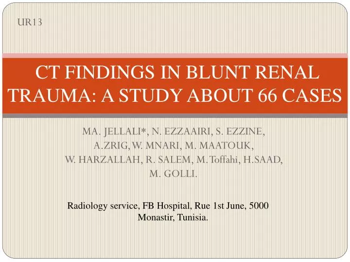
UR13. CT FINDINGS IN BLUNT RENAL TRAUMA: A STUDY ABOUT 66 CASES. MA. JELLALI*, N. EZZAAIRI, S. EZZINE, A.ZRIG, W. MNARI, M. MAATOUK, W. HARZALLAH, R. SALEM, M. Toffahi , H.SAAD, M. GOLLI. Radiology service, FB Hospital , Rue 1st June, 5000 Monastir, Tunisia . INTRODUCTION :.

E N D
UR13 CT FINDINGS IN BLUNT RENAL TRAUMA: A STUDY ABOUT 66 CASES MA. JELLALI*, N. EZZAAIRI, S. EZZINE, A.ZRIG, W. MNARI, M. MAATOUK, W. HARZALLAH, R. SALEM, M. Toffahi, H.SAAD, M. GOLLI. Radiology service, FB Hospital, Rue 1st June, 5000 Monastir, Tunisia.
INTRODUCTION : • Blunt trauma of the kidney remains a hot topic because of its high frequency. • Renal injury is observed in about 10% of cases of abdominal injury. • However, the majority (80%–90%) of renal injuries are attributable to blunt trauma, while the rest are due to penetrating renal injuries. • Both kidneys are at equal disposition for injury.
Renal injuries from blunt trauma usually occur as a consequence of a direct blow to the flank or from rapid deceleration. • A direct blow crushes the kidney, causing a laceration or lacerations of the renal parenchyma and resulting in a subcapsular, intrarenal or perinephrichaematoma. • A deceleration injury causes an acute tension on the renal pedicle, resulting in the laceration of the renal vein or artery, an intimal tear in the vessel causing thrombosis or laceration, or an avulsion of the ureteropelvic junction (UPJ).
All subsets of renal trauma require a high index of clinical awareness and prompt evaluation and management. • With the advancement in radiologic imaging modalities and the ready availability of computed tomography, the approach to renal injuries has changed over time, requiring diligent attention to recent literature. • Namely, the tolerance for nonoperative or expectant management has increased, even in the most seriously injured kidneys, replacing the past tendency toward aggressive renorrhaphy.
OBJECTIVES: • The objective of this study is to review the specific diagnosis of blunt trauma of the kidney with emphasis on the contribution of the Computed Tomography (CT) and especially to define the current position of conservative treatment.
MATERIALS AND METHODS: • The characteristics of this disease were reviewed from a retrospective study of 66 kidney injuries collected in the urology department of EPS F. Bourguiba in Monastir for a period of 15 years. • The assessment of the lesions was based primally on CT.
RESULTS: • Patients were predominantly male with a mean age of 25 years. • The causes were largely dominated by road accidents (41%). • The clinical presentation was dominated by low back pain (77%) and haematuria (59%). • The trauma was associated with abdominal visceral injury for 17 patients.
Was used the classification of Federle and our patients were as follows: 28 cases stage I, 17 cases stage II, 18 cases stage III, and 3 cases stage IV. • Four patients underwent emergency surgery for uncontrollablehemorrhage (intraabdominallesionsassociated). Fortythree patients (65%) withminor trauma(stage I and II) underwent non operative conservative treatment. Ten patients (15%) underwentdelayed five of themhad a total nephrectomy.
GRADE I SUBCAPSULAR HAEMATOMA RENAL CONTUSION
GRADE II LEFT RENAL LACERATION THROUGH THE CORTEX WITHOUT EXTENDING THE MEDULLA
GRADE IV LACERATION EXTENDING THE COLLECTING SYSTEM GRADE III LACERATION OF THE LEFT KIDNEY WITH AN HEPATIC CONTUSION
RELEVANT ANATOMY: • In most instances, the kidneys are paired retroperitoneal structures. They lie against the psoas muscles. • The superior aspect of the kidneys is somewhat protected by the lower ribs. However, the lower poles are inferior to the 12th ribs. • The parenchyma of the kidney has a segmental arterial supply. • This anatomic arrangement becomes important in the management of renal lacerations.
Blunt injuries tend to fracture along the planes between the segmental vessels, while penetrating injuries cross the segmental vessels. • Numerous anatomic variations exist, including pelvic kidneys; horseshoe kidneys; and multiple renal arterial, venous, and ureteral duplications.
EPIDEMIOLOGY: The frequency of renal injury somewhat depends on the patient population being considered. Renal trauma accounts for approximately 3% of all trauma admissions and as many as 10% of patients who sustain abdominal trauma.
PRESENTATION: The diagnosis of renal injury begins with a high index of clinical awareness. The mechanism of injury provides the framework for the clinical assessment. Particular attention should be paid to complaints of flank or abdominal pain. Urinalysis, both gross and, if necessary, microscopic, should be performed in patients who are thought to have renal trauma. Based on these initial measures, radiographic or operative investigation may follow.
DISCUSSION: • Given the high rate of renal loss and the improved results with expectant treatment, the decision to perform immediate surgery must be weighed carefully. • In decision making process, accurate assessment of the extent of injury is invaluable. • Intravenous urography (IVU) was previously the imaging modality of choice in the investigation of renal injuries.
However, it has been noted that IVU fails to detect and accurately stage some types of renal injuries. • Ultrasonography (US) has also been found to be very useful in the early evaluation of renal trauma, especially in the emergency room, as well as for detecting haemoperitoneum. However, US may also miss some types of renal injuries. • Computed tomography (CT) is currently the imaging modality of choice in the evaluation of blunt renal injury.
It can provide the exact delineation and staging of the extent of the renal injuries, and is superior to IVU, US and angiography. • However, CT needs to be performed in multiple phases for a complete assessment of renal injuries. • In certain cases, a delayed CT may need to be repeated after 2–3 days in order to detect ureteropelvic injuries and other complications.
Use of CT staging of renal trauma is sufficiently accurate to allow the majority of the patients with major renal injuries to be treated expectantly and to avoid unnecessary exploration with its high risk of renal loss. • Unless immediate exploratory laparotomy is indicated because of associated injuries or shock, most major renal injuries can be managed by non-surgical treatment with delayed intervention as needed.
The indications for renalimaging in trauma patients include: • grosshaematuria, • microscopichaematuriawithshock (systolicblood pressure [SBP] < 90 mmHg), • microscopichaematuriawithflankbruising, lowerrib and lumbarspine transverse process fractures, • penetrating trauma, and • a childwithblunt trauma and haematuria (> 50 redbloodscells/high power field).
CT SCANNING PROTOCOL IN RENAL TRAUMA: For a complete assessment of renal injuries, CT is performed in multiple phases. It is usually done as part of the CT abdomen and pelvis protocol for abdominal injuries. The corticomedullary phase is performed from the dome of the diaphragm to the pelvis, approximately 60 seconds after an intravenous injection of nonionic iodinated contrast media (iohexol 300 mg I/ml) at 2 mg/kg via the antecubital vein.
This phase would identify any renal contusion, laceration, perinephrichaematoma and arterial injury. Other associated injuries to the liver, spleen, pancreas and intraperitonealhaemorrhage could also be assessed. However, collecting system injuries may be missed if an excretory phase is not performed. The excretory phase is then obtained about 3–5 minutes later, which include both the kidneys and the urinary bladder. This is important in detecting urine extravastion which would indicate collecting system, ureteropelvic or bladder injuries.
The timing of the excretory phase may be delayed until more than 10–20 minutes so as to allow more urine extravasation to be visualised. In haemodynamically unstable patients, or patients with Category II or higher injuries, CT of the abdomen may need to be performed 2–3 days later to detect delayed complications, such as urinoma, infected urinoma or expanding haematoma, which may require intervention. All these multiphasicimagings of the renal system allow for appropriate and complete assessment of the renal injuries.
CT CLASSIFICATION OF RENAL TRAUMA: • There are several classifications of renal injuries, based on either imaging or surgery. Federle Classification is a widely used imaging-based grading system (Table I),while the American Association for the Surgery of Trauma (AAST) renal injury severity scale is a commonly used urological surgical staging in renal trauma (Table II). • However, considerable overlapping is observed in both classifications.
Staging is important as it guides surgeons and radiologists in the management of the patient, although it is not absolute and needs to be tailored for individual patients. • Out of the 66 cases that we have identified, 28 (42%) cases were in Category I (minor injury), 17 (25%) were in Category II (major injury), 18 (27%) were in Category III (catastrophic injury) and 3 cases (0.5%) were in Category IV (ureteropelvic junction injury).
Table I. Federle Classification (imaging-based) Category Type Injury I Minor injuryRenal contusion; intrarenal and subcapsularhaematoma; minor lacerationwithlimitedperinephric haematomawithout extension in the collecting system or medulla; small subsegmental cortical infarct II Major injury Major renal laceration through the cortex extending to the medulla or collecting system with or without urine extravasation; segmental renal infarct III Catastrophic injury Multiple renal lacerations; vascular injury involving the renal pedicle IV Ureteropelvic injury Avulsion (complete transaction); laceration (incompletetear)
Table II. The American Association for the Surgery of Trauma (AAST) renal injury severity scale: Grade Type Description I Contusion Microscopic or gross haematuria; urological studies normal HaematomaSubcapsular; not expanding with no parenchymal laceration II Haematoma Not expanding perirenalhaematoma confirmed to be renal retroperitoneum Laceration < 1.0 cm parenchymal depth of renal cortex with no urinary extravasation III Laceration > 1.0 cm parenchymal depth of renal cortex with no collecting system rupture or urinary extravasation IV Laceration Parenchymal laceration extending through renal cortex, medulla and collecting system Vascular Main renal artery or vein injury with contained haemorrhage V Laceration Completely shattered kidney Vascular Avulsion of renal hilum which devascularises kidney
Graphical representation of the American Association of Surgeons in Trauma grading system
CONCLUSION: Contrast- enhanced CT is readily available in emergency departments and can quickly and accurately depict renal injuries as well as associated injuries to other abdominal or retroperitoneal organs. It can provide essential anatomic and physiologic information required to determine management of injuries sustained during blunt abdominal trauma.