Download
1 / 75
790 likes | 1.13k Views
Oncologic Emergencies. Jim Holliman, M.D., F.A.C.E.P. Professor of Military and Emergency Medicine Uniformed Services University of the Health Sciences Clinical Professor of Emergency Medicine George Washington University Bethesda, Maryland, U.S.A. Oncologic Emergencies

E N D
Oncologic Emergencies Jim Holliman, M.D., F.A.C.E.P. Professor of Military and Emergency Medicine Uniformed Services University of the Health Sciences Clinical Professor of Emergency Medicine George Washington University Bethesda, Maryland, U.S.A.
Oncologic Emergencies Introduction • Malignancy is 2nd leading cause of death in U.S. • Now cancer has 52 % 5 year survival overall • Rx of complications can be life-saving since causative tumor often is curable • Rx of complications can, at a minimum, improve quality of life
List of Major Emergency Complications of Malignancy • Upper airway obstruction • Malignant pericardial tamponade • Superior vena cava syndrome • Acute spinal cord compression • Hypercalcemia • Hyperviscosity syndrome • Hyperleukocytic syndrome • Acute tumor lysis syndrome • SIADH • Adrenal insufficiency / crisis • Thrombocytopenia / hemorrhage • Immunosuppression / infection
Upper Airway Obstruction by Malignancy • Causative tumors : • Laryngeal ca • Thyroid ca • Lymphoma • Metastatic lung ca • Retropharyngeal abscess
Upper Airway Obstruction by Malignancy • Symptoms • Voice change • Hoarseness • Neck fullness • Dysphagia • Stridor • Dyspnea • Usually progresses & presents subacutely, unless food aspiration, infection, hemorrhage, or inspissated secretions occur
Upper Airway Obstruction by Malignancy • Diagnosis • Lateral soft tissue neck film • CXR • Fiberoptic laryngoscopy
Upper Airway Obstruction by Malignancy • Treatment • Oxygen • Racemic epinephrine aerosol (1.0 to 1.5 cc) • Helium / oxygen (Heliox) inhalation • ? IV steroids or diuretics • Intubation over fiberoptic laryngoscope • Consider tracheostomy • ? emergency radiation Rx
Malignant Pericardial Tamponade • Causative tumors : • Melanoma • Hodgkin's lymphoma • Acute leukemia • Lung ca • Breast ca • Ovarian ca • Radiation pericarditis • Rare to be initial presentation of malignancy
Malignant Pericardial Tamponade • Sx and Signs : • Dyspnea / weakness +/- chest pain • Hypotension / narrow pulse pressure • Friction rub rare • Jugular venous distention • Muffled (decreased) heart tones • Pulsus paradoxicus > 10 mm Hg • Low EKG QRS voltage +/- pulsus alternans • +/- cardiomegaly on CXR
Malignant Pericardial Tamponade • Dx : • Echocardiography • Equalization of heart chamber pressures • Rx options : • Needle catheter pericardiocentesis • Pericardial window under local anesthesia • Radiation Rx • Pericardiectomy • Intrapericardial chemoRx or sclerosis
Septum Posterior ventricular wall Pericardial effusion M mode echocardiogram showing malignant pericardial effusion
Two dimensional echo in same patient showing pericardial effusion (P)
Superior Vena Cava (SVC) Syndrome • Causative tumors : • Small cell (oat cell) lung ca • Squamous cell lung ca • Lymphoma • Anaplastic mediastinal ca • SVC thrombosis from indwelling catheter • Sx are due to SVC compression or occlusion
SVC Syndrome SYMPTOM FREQUENCY (%) Dyspnea 83 Cough 70 Orthopnea 64 Nasal congestion 35 Hoarseness 35 Stridor 33 Dizziness 29 Stupor / coma 20 Other sx : syncope, headache, dysphagia, epistaxis
SVC Syndrome SIGN FREQUENCY (%) Neck vein distention 92 Facial swelling / fullness 86 Arm vein distention 68 Mentation changes 27 Tongue edema 24 Laryngeal edema 24 Rhinorrhea 18
SVC Syndrome • Less common signs : • Facial plethora / telangiectasia • Supraclavicular palpable mass • Horner's syndrome • Papilledema • If present, represents a true emergency
SVC Syndrome • Diagnosis • CXR abnormal in 84 % • Confirm with (one of ) : • Chest CT with contrast • MRI • Contrast venography • Tc99m radionuclide venography
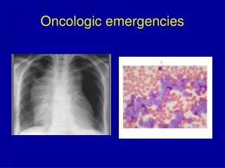
Mediastinal widening and pulmonary venous obstruction from lung cancer
Chest computed tomography showing right sided mediastinal lung cancer compressing the contrast filled SVC
Another CT cut of the same patient showing tumor compression of the SVC
37 year old male who presented with sudden onset of venous stasis, hoarseness, and hemoptysis
Chest X-ray of same patient who proved to have squamous cell cancer of the lung blocking the left subclavian vein
SVC Syndrome • Treatment : • Keep in head-up position • IV steroids • IV diuretics • ? anticoagulants or thrombolytics • Emergent mediastinal radiation Rx • Remove central IV catheter if present
Acute Spinal Cord Compression • Causative tumors : • Breast ca • Lung ca • Prostate ca • Lymphomas • Multiple myeloma • Renal cell ca • Sarcomas • Epidural abscess / hematoma • 18,000 cases per year in U.S.
Acute Spinal Cord Compression • Symptoms : • Localized back pain +/- tenderness • May be absent with lymphomas • Paraparesis / paraplegia • Distal sensory deficits • Urinary incontinence
Acute Spinal Cord Compression • Cervical, thoracic, or lumbar spine films : 85 % abnormal • May not be needed if CT or MRI planned anyway • Radionuclide bone scan • Sensitivity > 90 % except for multiple myeloma • Spine CT with contrast • MRI • Myelography • NOTE : any studies done should be in emergent time frame & with early involvement of consultant
MRI scan showing multiple myeloma destroying the C6 vertebra and surrounding the spinal cord
40 year old male who presented with neck pain ; he had a metastatic hypernephroma which destroyed the odontoid
Myelogram showing breast cancer metastasis pressing on the L5 nerve root, and also pushing the thecal sac posteriorly at L4
T1 MRI of same patient showing compression of the L5 nerve root
Acute Spinal Cord Compression • Treatment • Spine immobilization • Foley catheter • ? IV steroids / diuretic / mannitol • Emergent decompressive laminectomy or radiation Rx
Hypercalcemia of Malignancy • Causative tumors • Metastatic breast, lung, or prostate ca • Multiple myeloma • Non-Hodgkin's lymphoma • Adult T-cell lymphoma / leukemia • Renal cell ca • Head & neck squamous cell ca
Malignancy Hypercalcemia • Symptoms • Vague malaise / weakness • Polydipsia • Lethargy / confusion • Constipation • Vomiting • Back pain • Can have coma or seizures
Malignancy Hypercalcemia • Diagnosis • Total & ionized serum calcium • Serum albumin sometimes helpful • EKG shows short QT interval • May show low voltage, long PR • Discrete skeletal lesions not demonstrable in 30 % of patients • Serum levels > 12 mg % dangerous
EKG showing short QT interval (0.28 seconds) in a patient with a serum calcium of 14 mg/dl
Malignancy Hypercalcemia Treatment • IV hydration with normal saline • Diuresis with IV furosemide • Only after fluid loading ; avoid thiazides • IV steroids • Etidronate (7.5 mg/kg/day IV for 3 days) • Mithramycin (15 to 25 mcg/kg/day IV x 3 days) • Radiation Rx to tumor site(s) • Rarely may need hemodialysis
Considerations for Use of Etidronate (Didronel) for Rx of Malignant Hypercalcemia • Acts mainly to reduce bone resorption • Mainly excreted renally • Causes some degree of hyperphosphatemia • Should be withheld if creatinine > 5 mg % • Dose (must be diluted in 250 cc NS) : • 7.5 mg/kg/day IV for 3 days • Dose should be given over 2 hours • Followup Rx with oral tablets • 20 mg/kg/day for 30 days
Use of Mithramycin (Plicamycin) for Rx of Malignancy Hypercalcemia • Acts as antineoplastic agent • Method of action on hypercalcemia not known • Main complication is bleeding • GI side effects common • Can cause thrombocytopenia • Most useful as second agent for cases not responsive to etidronate
Hyperviscosity Syndrome • Basic cause is elevation of serum proteins producing sludging & reduction in microcirculatory perfusion • Serum viscosity is normally 1.4 to 1.8 times that of water • Symptoms develop at viscosity > 5
Hyperviscosity Syndrome • Causative tumors • Multiple myeloma • Waldenstrom's macroglobulinemia • Chronic myelocytic leukemia
Hyperviscosity Syndrome Symptoms • Fatigue / malaise • Headache • Anorexia • Somnolence • If microthromboses occur : • Deafness • Visual deficits • Seizures