Download
1 / 26
270 likes | 297 Views
CARDIAC ELECTRIC ACTIVITY: CONDUCTING SYSTEM. Prof. Sultan Ayoub Meo MBBS, M.Phil, Ph.D (Pak), M Med Ed (Dundee), FRCP (London), FRCP (Dublin), FRCP (Glasgow), FRCP (Edinburgh) Professor and Consultant, Department of Physiology, College of Medicine, King Saud University, Riyadh, KSA.

E N D
CARDIAC ELECTRIC ACTIVITY: CONDUCTING SYSTEM Prof. Sultan Ayoub Meo MBBS, M.Phil, Ph.D (Pak), M Med Ed (Dundee), FRCP (London), FRCP (Dublin), FRCP (Glasgow), FRCP (Edinburgh) Professor and Consultant, Department of Physiology, College of Medicine, King Saud University, Riyadh, KSA
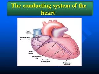
COMPONENTS OF CONDUCTIVE SYSTEM • S A Node • Inter-Nodal Pathway • A V Node • A V bundle • Right bundle branch • Left bundle branch
CONDUCTING TISSUES OF THE HEART Heart has a special system for generating rhythmical electrical impulses to cause rhythmical contraction of the heart muscle.
Sinus-Atrial Node (SA node) Atria Atrial-ventricular Node (AV node) Ventricles Sequence of excitation CONDUCTING TISSUES OF THE HEART
CONDUCTING TISSUES OF THE HEART Sequence of excitation
SA NODE • The Sinus Node (Sinoatrial node) is a small, flattened, specialized cardiac cell • Known as Pacemaker of the heart. • Located in the superior posterio-lateral wall of the right atrium • Responsible for generating the electrical impulses that bring about the mechanical activity i.e contraction of the heart. • SA node has the fastest rate of autorhythmicity.
CARDIAC IMPULSE FROM SA NODE TO ATRIAL MUSCLE The cardiac impulse after it’s origin in the SA node spreads through out the atrial muscle through two routes • Ordinary Atrial muscle fibers • Anterior, middle and posterior conducting bundles • Anterior internodal bundle of Bachman • Middle internodal bundle of Wenkebach • Posterior internodal bundle of Thoral • These inter nodal pathways conduct the impulses at a faster rate than atrial muscle fibers, because of specialized conduction fibers.
CARDIAC IMPULSE FROM SA NODE TO ATRIAL MUSCLE • The velocity of conduction in most atrial muscle is about 0.3m/sec. • In the specialized internodal pathways the conduction velocity may reach upto 1m/sec. • The impulse after leaving SA node takes 0.03 sec to reach the AV node.
AV NODE The AV node is located in the posterior wall of the right atrium immediately behind the tricuspid valve. Slow Conduction in the AV Node: The cause of slow conduction is mainly diminished number of gap junctions between the successive cells in the conducting pathways.
SIGNIFICANCE OF AV NODAL DELAY • The cardiac impulse does not travel from the atria to the ventricles too rapidly. • This delay allows time for the atria to empty their blood into the ventricles before ventricular contraction begins. • This increases the efficiency of the pumping action of the heart.
PURKINJEE FIBERS • Purkinje fibers are very large fibers and they transmit AP at a velocity of 1.5 to 4.0 m/sec. • The rapid transmission of action potentials through the Purkinje fibers is believed to be caused by a very high level of permeability of gap junctions at the intercalated discs between the successive cells of Purkinje fibers. • The rapid conduction through the purkinje fibers ensures that different parts of ventricles are excited almost simultaneously; this greatly increases the efficiency of heart as a pump.
RIGHT AND LEFT BUNDLE BRANCHES • Bundle of His splits into two branches which are called right and left bundle branches present on the respective sides of the ventricular septum. • From the time the cardiac impulse enters the bundle branches until it reaches the terminations of Purkinje fibers , the total time averages only 0.03 sec.
CONDUCTION OF IMPULSE • APs from SA node spread quickly at rate of 0.8 - 1.0 m/sec. • Time delay occurs as impulses pass through AV node. • Slow conduction of 0.03 – 0.05 m/sec. • Impulse conduction increases as spread to Purkinje fibers at a velocity of 4.0 m/sec. • Ventricular contraction begins 0.1–0.2 sec. after contraction of the atria.
ONE- WAY CONDUCTION THROUGH AV BUNDLE • A special characteristic of the A-V bundle is it’s inability of action potentials to travel backward from the ventricles to the atria. • This prevents re-entry of cardiac impulse by this route from the ventricles to the atria. • The atrial muscle is separated from the ventricular muscle by a continuous fibrous barrier which acts as an insulator to prevent the passage of cardiac impulse between the atrial and ventricular muscle
ACTION POTENTIAL IN THE CARDIAC MUSCLE The cardiac action potential is made of 3 phases: Depolarization: caused by the opening of Fast Na channels & slow Ca channels Plateau: remaining of slow Ca channels open for several m seconds, drawing large amount of Ca inside which prolong depolarization Replarization: Opening of potassium channels
ACTION POTENTIAL IN THE CARDIAC MUSCLE The presence of Plateau in the action potential causes ventricular contraction to last as much as 15 times as long in cardiac muscle as in skeletal muscle Guyton pp 110
ACTION POTENTIAL IN THE CARDIAC MUSCLE Phase 0 (depolarization): Fast sodium channels open. Voltage gated sodium channels (fast sodium channels) open and cell depolarize. Membrane potential reaches about +20 millivolts before the sodium channels close. Phase 1 (initial repolarization), fast sodium channels close. Cell begins to repolarize, and potassium ions leave the cell through open potassium channels. Guyton and Hall, pp 111
ACTION POTENTIAL IN THE CARDIAC MUSCLE Phase 2 (Plateau), calcium channels open and fast potassium channels close. Initial repolarization Occurs. Potassium ion efflux and increased calcium ion influx causes the action potential to plateau. Phase 3 (rapid repolarization), calcium channels close and slow potassium channels open. The closure of calcium ion channels and increased potassium ion permeability, permitting potassium ions to rapidly exit the cell Phase 4 (resting membrane potential) averages about −90 millivolts. Guyton and Hall, pp 111
WHY PLATEAU OCCURS • Voltage activated sodium channels, called fast channels • Voltage-activated calcium-sodium channels (L-type calcium), slow to open, called slow channels. • Opening of fast channels causes spike of AP • Prolonged opening of the slow calcium-sodium channels allows calcium to enter, cause plateau. • Moreover, voltage-gated potassium channels are slower to open. This delays the return of the membrane potential to −80 to −90 millivolts.
Refractory Periods • Heart contracts as syncytium. • Contraction lasts almost 300 msec. • Refractory periods last almost as long as contraction. • Myocardial muscle cannot be stimulated to contract again until it has relaxed. • Summation cannot occur.
Long refractory period prevent ventricles from contracting at too high rates so that enough time is allowed for refill of the ventricles • Because long refractory period occurs in conjunction with prolonged plateau phase, summation and tetanus of cardiac muscle is impossible • Ensures alternate periods of contraction and relaxation which are essential for pumping blood
Conduction Cardiac Muscle Velocity of Signal Conduction in Cardiac Muscle The velocity of conduction of the excitatory action potential in atrial and ventricular muscle fibers is about 0.3 to 0.5 m/sec, or about 1/250 the velocity in very large nerve fibers and 1/10 the velocity in skeletal muscle fibers. The velocity of conduction in the specialized heart conductive system in the Purkinje fibers is as great as 4 m/sec