Download

1 / 73
730 likes | 931 Views
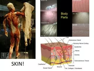
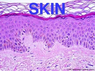
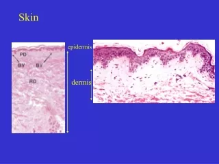
Skin. Object is for you to learn and identify skin lesions. Describe his skin. Normal skin. Color: Brown Normal hair No lesions Should feel for texture, turgor, moisture and warmth. Describe the lesion. Normal Mole. Tan to brown Uniformly pigmented

E N D
Skin Object is for you to learn and identify skin lesions
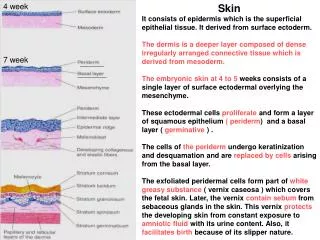
Normal skin Color: Brown Normal hair No lesions Should feel for texture, turgor, moisture and warmth
Normal Mole • Tan to brown • Uniformly pigmented • Small (usually less than 6 mm across) • Solid regions of relatively flat (macules) to elevated skin (papules) • Well-defined, rounded borders
Chronic ITP Hyperpigmentation
Campbell’d morgan spots Cherry angiomas are • Bright red • Small (usually 1-4 mm) • Papules • Commonly seen on the trunk of adults. • Medically insignificant
Hodgkin’s disease • Dry • Scaly
Senile purpura • Red • Patch • Does not blanch on pressure • Thin skin
Steroid induced purpura • Red • Patch • Does not blanch on pressure
Schonlein-Henoch purpura • Ankle locaton • Multiple • Red • Not blanching on pressure • Less than 5 mm • Vasculitis
Henoch Schonlein purpura • Multiple • Red • Not blanching on pressure • Less than 5 mm • Vasculitis
Echymosis • Red • Large patch • Irregular edge • Changing colors Coagulation defect
Vasculitis • Multiple • Red • Not blanching on pressure • Less than 5 mm • Coalesing Vasculitis drug induced
Leukemia cutis • Ulcerating lesions Black base Sharp edges Surrounding echymoses • Blister 1 cm Fluid filled
Kaposi Sarcoma • Nonblanching red macule • Surrounding ecchymoses and acquire more of a violet hue • The lesions may become nodular
Malignant Melanoma A: asymmetry- one side or half does not look like the other B: border irregularity C: color-black, or much darker than patient's other moles, often with red, white or blue areas. D: diameter > 6 millimeters
Sarcoidosis • Patch • O.5-1 cm in size • Different color than skin • Slightly elevated Maculo-papular lesions
Sarcoidosis • Multiple • Less than 1 cm • Raised spot • Dome shaped Papular lesions
Sickle cell anemia • Over ankle • Patch • larger than 1 cm • Thin skin • Loss of substance of skin Old scar
Normal Transluscent Shiny Appears firm
Doxorubicin • Transeverse band • White in color
Iron deficiency anemia • Spooning • concave Koilonychia
Ehler’s danlos syndrome • Patch • larger than 1 cm • Thin skin • Loss of substance of skin Old widened scar
Gardner Diamond syndrome • Multiple • Leniar wide • Thin skin Striae
Suqmous cell carcinoma • Ulcer • Ear lobe • Crusted lesion • Sharp margin • Indurated
Keloid (after piercing) • Very firm • Rubbery lesions • Reddish or darkly colored • Occur after trauma sometimes very minor trauma • May itch.