Download
1 / 7
70 likes | 175 Views
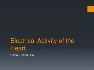
Your Electrical Heart. Exploring EKG. Objectives. Find and interpret patterns on an EKG graph Describe the electrical and mechanical components of a normal heart rhythm Draw appropriate conclusions about the mechanics of various normal and abnormal heart functions based on EKG. Introduction.

E N D
Your Electrical Heart Exploring EKG
Objectives • Find and interpret patterns on an EKG graph • Describe the electrical and mechanical components of a normal heart rhythm • Draw appropriate conclusions about the mechanics of various normal and abnormal heart functions based on EKG
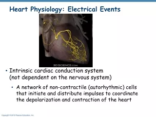
Introduction • When have you heard the terms “V-tac”, “V-fib”, “asystole”? • What is an EKG or ECG? • What do you think a normal graph of the heart’s electrical activity looks like? (draw it) • Why do doctors and nurses need EKGs? Depiction of electrical activity in the heart Detect & diagnose problems with the patient’s heart rhythm
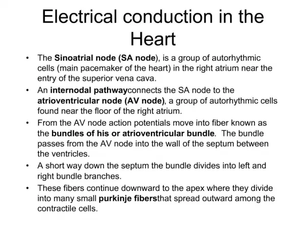
Review and new terms • Left & right atria: receive blood from the veins, pump it into ventricles • Left & right ventricles: receive blood from atria, pump it out into arteries • Sinoatrial (SA) node: bundle of nerves in R atrium, the heart’s pacemaker, stims contraction of atria • Atrioventricular (AV) node: between atria & ventricles, continues rhythm set by SA node, stims contraction of ventricles • Purkinje fibers: fan out and stimulate contraction of ventricles
Review: the cardiac cycle • How blood moves through the heart • Blood enters atria>>pressure in A rises>>AV valves open • Blood enters ventricles>>pressure in V rises>>AV valves close>>semilunar valves open>>blood leaves heart • Pressure in V falls>>S/L valves close
To Do and Notice • Look at your EKG strip. • Determine the patient’s heart rate in BPM if this is a 6 second rhythm strip. • Is the rhythm consistent? (describe) • How is your strip similar to a normal EKG? How is it different? • Based on what you know about the cardiac cycle, try to explain what is going wrong with this heart. • Present your patient to the class.
What’s Going On? • What terms and conditions do we learn from the EKGs?