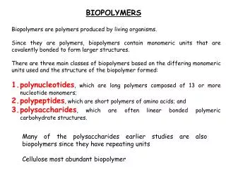
Download

1 / 31
310 likes | 491 Views
Spectroscopy of Biopolymers. Protein Function. Functions of Proteins. Binding

E N D
Spectroscopy of Biopolymers Protein Function
Functions of Proteins • Binding The most fundamental of these is binding, which underlies all the other biochemical functions of proteins. Enzymes must bind substrates, as well as cofactors that contribute to catalysis and regulatory molecules that either activate or inhibit them. • Catalysis • Switching • Structural Proteins assemblages of a single type of protein molecule bound together for strength or toughness; in more complex cases they bind to other types of molecules to form specialized structures such as the actin-based intestinal microvilli or the spectrin-based mesh that underlies the red blood cell membrane and helps maintain its integrity as the cells are swept round the body.
Binding • Specific recognition of other molecules is central to protein function. Myoglobin binds a molecule of oxygen reversibly to the iron atom in its heme group (shown in grey with the iron in green). It stores oxygen for use in muscle tissues. The TATA binding protein binds a specific DNA sequence and serves as the platform for a complex that initiates transcription of genetic information.
Catalysis • Essentially every chemical reaction in the living cell is catalyzed, and most of the catalysts are protein enzymes. • The catalytic efficiency of enzymes: reactions can be accelerated by as much as 17 orders of magnitude over simple buffer catalysis. • Many structural features contribute to the catalytic power of enzymes: holding reacting groups together in an orientation favorable for reaction (proximity); binding the transition state of the reaction more tightly than ground state complexes (transition state stabilization)
DNA replication is catalyzed by a specific polymerase that copies the genetic material and edits the product for errors in the copy. Replication of the AIDS virus HIV depends on the action of a protein-cleaving enzyme called HIV protease. This enzyme is the target for protease-inhibitor drugs (shown in grey).
Switching Proteins are flexible molecules and their conformation can change in response to changes in pH or ligand binding. Such changes can be used as molecular switches to control cellular processes. The GDP-bound ("off"; PDB 1pll) state of Ras differs significantly from the GTP-bound ("on"; PDB 121p) state.This difference causes the two states to be recognized by different proteins in signal transduction pathways.
Structural Proteins Protein molecules serve as some of the major structural elements of living systems. This function depends on specific association of protein subunits with themselves as well as with other proteins, carbohydrates, and so on, enabling even complex systems like actin fibrils to assemble spontaneously. Structural proteins are also important sources of biomaterials, such as silk, collagen, and keratin. Silk derives its strength and flexibility from its structure: it is a giant stack of antiparallel beta sheets. Its strength comes from the covalent and hydrogen bonds within each sheet; the flexibility from the van der Waals interactions that hold the sheets together. Actin fibers are important for muscle contraction and for the cytoskeleton. They are helical assemblies of actin and actin-associated proteins.
Tubulin (a) The biochemical functions of tubulin include binding of tubulin monomers to each other to form a polymeric protofilament, a process that is reversed by the hydrolysis of bound GTP to GDP. Tubulin-catalyzed hydrolysis of GTP actsas a switching mechanism, in that rotofilaments in the GDP form rapidly depolymerize unless the concentration of free tubulin is very high or other proteins stabilize them. (b) This nucleotide-dependent mechanism is used by the cell to control the assembly and disassembly of the protofilaments and the more complex structures built from them. These structures include microtubules, which consist of 13 protofilaments arranged as a hollow tube (here shown in growing phase). (c) Binding to motor proteins such as kinesin or dynein allows the microtubules to form molecular machines in which these motor proteins “walk” along microtubules in a particular direction, powered by ATP hydrolysis.
Tubulin The functions of these machines are defined at the cellular level. Assemblies of microtubules, motor proteins and other microtubule-associated proteins form the flagella that propel sperm, for example (d); microtubules and associated motor proteins also form a network of “tracks” on which vesicles are moved around in cells (e). The “role” of tubulin thus encompasses both biochemical and cellular functions. The individual functions of proteins work in concert to produce the exquisite machinery that allows a cell, and ultimately a multicellular organism, to grow and survive. The anti-cancer drug taxol blocks one essential cellular function of tubulin. It binds to the polymerized protein, preventing the disassembly of microtubules that must occur during cell division.
Recognition and Complementarity • Function and Flexibility
Recognition and Complementarity • Protein functions such as molecular recognition and catalysis depend on complementarity. • Molecular recognition depends on specialized microenvironments that result from protein tertiary structure. • Specialized microenvironments at binding sites contribute to catalysis.
Recognition and Complementarity • The functions of all proteins, whether signaling or transport or catalysis, depend on the ability to bind other molecules, or ligands. • The ligand that is bound may be a small molecule or a macromolecule, and binding is usually very specific. • Ligand binding involves the formation of noncovalent interactions between ligand and protein surface. • Specificity arises from • the complementarity of shape and charge distribution between the ligand and its binding site on the protein surface. • the distribution of donors and acceptors of hydrogen bonds.
Substrate binding to anthrax toxin lethal factor Lethal factor (LF) is a component of anthrax toxin that acts as a protease to cut mitogen-activated protein kinase kinase (MAPKK-2), thereby blocking the cell cycle. This figure shows part of the surface of LF colored by charge (red, negative; blue, positive), with the model of the MAPKK-2 amino-terminal peptide shown in ball-and-stick representation. Where the model or map would be hidden by the protein surface, the surface is rendered as translucent. The active-site cleft of LF is complementary in shape and charge distribution to the substrate.
Flexibility and Protein Function • The flexibility of tertiary structure allows proteins to adapt to their ligands. • Protein flexibility is essential for biochemical function. • The degree of flexibility varies in proteins with different functions.
Flexibility and Protein Function The flexibility of tertiary structure allows proteins to adapt to their ligands. Tight fit between a protein and its ligand catalytic domain of protein kinase A (blue) bound to a peptide analog (orange) of its natural substrate shows the snug fit between protein and ligand, achieved by mutual adjustments made by the two molecules.
Flexibility and Protein Function HIV protease, an enzyme from the virus that causes AIDS, bound to three different inhibitors The anti-viral action of some drugs used in AIDS therapy is based on their ability to bind to the active site of viral protease and inhibit the enzyme. The protease inhibitors haloperidol (a) and crixivan (b) are shown, with a peptide analog (c) of the natural substrate also shown bound to the enzyme. Each inhibitor clearly has a quite different structure and two of them (a, b) are not peptides, yet all bind tightly to the active site and induce closure of a flap that covers it, a conformational change that also occurs with the natural substrate.
The enzyme adenylate kinase can adopt either an open or closed conformation depending on which substrates are bound. (b) On binding of the cosubstrate ATP, here in the form of the analog AMPPNP, a large rearrangement occurs that closes much of the active site. (a) In the presence of AMP alone, no conformational change occurs.
Active site and Binding site • active site: asymmetric pocket on or near the surface of a macromolecule that promotes chemical catalysis when the appropriate ligand(substrate) binds. • ligand-binding site: site on the surface of a protein at which another molecule binds.
Location of Binding Site • Binding sites for macromolecules on a protein’s surface can be concave, convex, or flat. • The most frequently observed sites are protruding loops or large cavities because these provide specific shape complementarity, but relatively flat binding sites are also found.
Growth Hormone The specific recognition of a macromolecule by a protein usually involves interactions over a large contiguous surface area (hundreds of square Å) or over several discrete binding regions (Figure 2-8). A macromolecule will make many points of contact with the protein’s surface. The complex between human growth hormone and two molecules of its receptor.
Many binding sites for RNA or DNA on proteins are protruding loops or alpha helices that fit into the major and minor grooves of the nucleic acid The complex between the bacterial diphtheria toxin gene repressor protein and the tox operator DNA sequence to which it binds. The repressor is a natural homodimer and two dimers bind to the pseudo-symmetrical operator sequence. The DNA sequence is recognized in the major groove by a helix in a helix-turn-helix motif, a feature often seen in DNA-binding proteins. Structure of the complex between the eukaryotic Gal4 transcription factor and DNA. The interaction occurs in the major groove again, but this time the recognition unit contains a loop of chain that is stabilized by a cluster of zinc ions.
Binding sites for small ligands are clefts, pockets or cavities Small-molecule ligands bind at depressions on the protein surface, except in certain cases when they are buried within the protein’s interior. Deep binding pockets allow the protein to envelop the ligand and thus use complementarity of shape to provide specificity. Clefts or cavities can easily provide unusual microenvironments.
camphor Heme group Binding in an interior cavity requires that the ligand diffuses through the protein structure within a reasonable time frame. Protein structural flexibility can allow such penetration, even for large substrates. Structure of bacterial cytochrome P450 with its substrate camphor bound
Catalytic sites often occur at domain and subunit interfaces NADPH Sub-unit Structure of the dimeric bacterial enzyme 3-isopropylmalate dehydrogenase
Nature of Binding Site • Binding sites generally have a higher than average amount of exposed hydrophobic surface. • Binding sites for small molecules are usually concave and partly hydrophobic. • Weak interactions can lead to an easy exchange of partners. • Displacement of water also drives binding events. • Contributions to binding affinity can sometimes be distinguished from contributions to binding specificity.
Ligand-binding sites are places where nonpolar groups tend to be clustered on the protein surface, and this physical-chemical characteristic can sometimes be used to recognize them. Surface view of the heme-binding pocket of cytochrome c6, with hydrophobic residues indicated in yellow. The area around theheme (red) is very nonpolar because this protein must bind to another protein via this site to form an electron-transport complex involving the heme. The blue area indicates the presence of two positively charged residues important for heme binding.
Functional Properties of Structural Proteins • Structural Proteins as frameworks, connectors and scaffolds • All cells are surrounded by a protein-reinforced membrane; some have a cell wall that is primarily protein and carbohydrate. • Internal structures within the cell also are made up of particular structural proteins that confer shape, strength and flexibility on these cellular structures. • In some cases, structural proteins are assisted by DNA, RNA, lipid and carbohydrate molecules; • in other cases the structure is built up from a large number of different proteins.
Structure of the 50S (large)subunit of the bacterial ribosome RNA The ribosome has over a hundred different protein components that stabilize the folded form of the ribosomal RNA, which provides the catalytic function
Some structural proteins only form stable assemblies The structural components of cells and organisms formed by proteins can be constructed from proteins alone, as in the case of silk, collagen, elastin or keratin, or the coat proteins of a virus or from protein plus some other component, as in the case of cartilage, which is composed of protein plus carbohydrate. • Structure can be stabilized in two ways : • Protein-protein interaction • Covalent cross-linking
Collagen, the fibrous component of tendons, is one example of such a structure: the basic component of collagen is a triple helix of three protein chains made of repeating GlyXY sequences. Collagen triple helix is a coiled-coil structure often proline (in the example shown here, Y is also proline). The hydrophobic nature of this repeat results in a set of regularly spaced hydrophobic sites along each chain; these complementary sites plus interchain hydrogen bonds (red dotted lines) hold the triple helix together. In collagen fibers, multiple triple helices are aligned end-to-end and side-by-side in a regular fashion, producing the light and dark bands observed when collagen fibers are imaged in an electron microscope. http://www.rcsb.org/pdb/molecules/pdb4_1.html
Some catalytic proteins can also have a structural role • The regular assemblies formed by structural proteins often have cellular functions that require time-dependent changes in shape or conformation or some other property of the assembly. These changes are often brought about by changes in the structure of a single component of the multicomponent assembly by binding of another protein or small molecule.