Download
1 / 27
320 likes | 658 Views
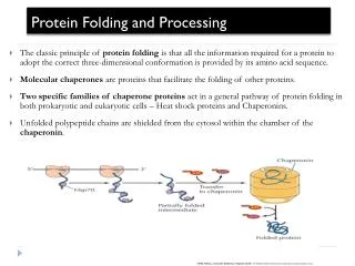
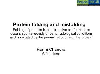
Protein expression is the multistep process involving regulation at the level of transcription , mRNA turnover , protein translation , and post-translational modifications leading to the formation of a stable product. Competing reactions of protein folding and aggregation.

E N D
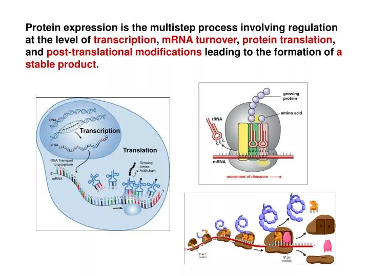
Protein expression is the multistep process involving regulation at the level of transcription, mRNA turnover, protein translation, and post-translational modifications leading to the formation of a stable product.
Competing reactions of protein folding and aggregation. Scheme of the free-energy surface that proteins explore as they move towards the native state (green) by forming intramolecular contacts. The ruggedness of the free-energy landscape results in the accumulation of kinetically trapped conformations that need to traverse free-energy barriers to reach a favourable downhill path. In vivo, these steps may be accelerated by chaperones.When several molecules fold simultaneously in the same compartment, the free-energy surface of folding may overlap with that of intermolecular aggregation, resulting in the formation of amorphous aggregates, toxic oligomers or ordered amyloid fibrils (red). Fibrillar aggregation typically occurs by nucleation-dependent polymerization. It may initiate from intermediates populated during de novo folding or after destabilization of the native state (partially folded states) and is normally prevented by molecular chaperones. NATURE | VOL 475 | 21 JULY 2011
Partially folded or misfolded states are problematic - they tend to aggregate in a concentration-dependent manner. Aggregation primarily results in amorphous structures. Alternatively, fibrillar aggregates called amyloid may form. Many proteins can adopt these thermodynamically stable structures under conditions in vitro, the formation of these aggregates in vivo is strongly restricted by the chaperone machinery. Molecular chaperone interact with, stabilize or help proteins to acquire its functionally active conformation, without being present in its final structure. Different classes of structurally unrelated chaperones exist in cells. Members of these protein families are often known as stress proteins or heat-shock proteins (HSPs), as they are upregulated under stress.
Amyloidní fibrila – patologická forma proteinu v beta struktuře (struktuře skládaného listu). Ke vzniku amyloidu dochází agregací původně solubilní (degradovatelné) formy proteinu do formy fibrilární (resistentní na degradaci). Stabilizace nastává především interakcí mezi jednotlivými polypeptidickými řetězci. Dochází tak, na rozdíl od alfa-helixu kde stabilizace probíhá uvnitř šroubovice, nejen k intramolekulární interakci mezi vzdálenými částmi polypeptidického řetězce, ale i k i intermolekulární agregaci mezi jednotlivými proteiny. Vzniklé fibrily se skládají z beta úseků, kde jednotlivé řetězce jsou navzájem uspořádány antiparalelně a leží kolmo na dlouhou osu vlákna. Fibrily jsou velmi resistentní na proteolysu a jejich degradace je tedy velmi omezená.
The nascent protein is synthesized vectorially and N-terminal sequences will be available for folding before the C-terminal fragments. • The rate of translation is coupled to the co-translational folding and can influence the final conformation of the protein. • The translation proceeds in a time scale of seconds (15–20 AA/s for prokaryotes and 1–5 AA/s for eukaryotes) and thus imposes an upper limit on the co-translational folding. • The rate of translation is non-uniform along mRNAs and is shaped by the asymmetric tRNA abundance for the different codons. Triplets that pair to lowly abundant tRNAs are translated slower that the codons read by highly abundant tRNA-species. Codons pairing to minor tRNAs tend to cluster forming within the mRNA slow-translating stretches at which ribosomes transiently attenuate the elongation cycle. • The slow-translating segments are not randomly distributed along the coding mRNA sequences; they are predominantly located downstream (usually 20–70 amino acids) of the domain boundaries of multidomain proteins. Precisely regulated ribosome speed, mediated by the codon choice, is crucial for accurate folding of the cognate protein. • Alterations in the pattern of slow-translating regions by synonymous substations to codons that are translated at different speed than the original triplet have a deleterious effect on the folding of multidomain proteins.
Clusters of codons pairing to minor tRNAs (red dots on the mRNA message) shape minima in the translation profile (red discontinuous line) causing the non-uniform traffic of the ribosomes along the mRNA. At these local minima the ribosomes transiently attenuate the elongation of the nascent peptide. Deep local minima kinetically coordinate the hierarchical folding of single domains in multidomain proteins, whereas less intensive minima may provide a kinetic window for formation of secondary structure. Current Opinion in Structural Biology 2011, 21:25–31
At the end of the last century, metabolic pediatricians recognized that some phenylketonuria patients may benefit from pharmacological doses of the natural cofactor tetrahydrobiopterin (BH4) of the deficient enzyme PAH. • The majority of patients with mild phenotypes benefited from cofactor treatment. BH4 reduced blood phenylalanine concentrations and increased dietary phenylalanine tolerance by increasing in vivo PAH activity. • In 2007 (FDA) and 2008 (EMEA), sapropterin dihydrochloride, the synthetic form of BH4, was approved as an orphan drug to treat patients with BH4-responsive PAH deficiency. • About 60% of all PKU patients are estimated to benefit from this new therapy. Patients with a mild to moderate clinical phenotype due to a genotype associated with some residual enzyme activity are more likely to respond to cofactor treatment than those with classic PKU carrying nonsense mutations • PKU - protein misfolding disease with loss of function. • BH4 - pharmacological chaperone. • A pharmacological chaperone is a small molecule that rescues protein function by improving protein folding and by stabilizing the protein structure.
Schematic view of the molecular pathophysiology of PAH deficiency. PAH mutations can lead to protein misfolding, aggregation, and early degradation and thus to a loss of functional PAH. All processes are mutually dependent and contribute to loss of function. J Inherit Metab Dis (2010) 33:649–658
Molecular mode of action of BH4. At the protein level, BH4 prevents misfolding, aggregation, and degradation, and thus induces an increase in the effective PAH concentration resulting in rescue of its function. J Inherit Metab Dis (2010) 33:649–658
Familialní hypercholesterolemie (FH) - mutace v genu pro low-density lipoprotein receptor (LDLR) nebo apolipoprotein B100 (APOB)- frekvence heterozygotů (1:500), frekvence homozygotů (1:1000000) Patogenní mutace v LDL receptoru způsobují ztrátu funkčních receptorů pro LDL částice na povrchu jaterních buněk → zvýšení hladiny LDL v plazmě → ateroskleróza a zvýšené riziko koronární srdeční nemoci
LDLR is synthesized by ribosomes bound to the endoplasmic reticulum (ER), partially glycosylated (molecular mass 120 kDa). • LDLR is transported to the Golgi apparatus, glycosylated (molecular mass 160 kDa). • LDLR is transported to the cell surface, where it mediates the uptake of lipoprotein particles, mainly low-density lipoproteins (LDLs), by receptor-mediated endocytosis. • The internalized LDL particle is subsequently released in the endosome, and the receptor returns to the cell surface in a process called receptor recycling. Cell. Mol. Life Sci. Vol. 61, 2004
The extracellular part of LDLR contains a series of discrete extracellular protein domains that are responsible for binding and release of LDL. • Ligand binding region - the N-terminal part; seven cysteine-rich repeats (~40 AA), which mediate the binding to LDL. • EGF-precursor homology region - two epidermal growth factor (EGF)-like modules (each ~40 AA), six-bladed b-propeller (~260AA), and third EGF-like module. This domain controls the LDL release at low pH in the endosomes and the recycling of the receptor to the cell surface. • O-linked sugar region - rich in serine and threonine residues, which undergo O-linked glycosylation. Domain organization of the LDL receptor Nature Structural Biology 8, 476 - 478 (2001)
Folding of LDLR has long been considered to occur in a vectorial manner, i.e. domain by domain, starting with the most N-terminal cysteine-repeat R1. • The newly synthesized LDLR chains fold rapidly into compact structures containing non-native disulfide bonds linking distant regions of the protein. • With time, the non-native disulfides are reshuffled (i. e. isomerized), allowing extension of the molecule. In the native conformation, disulfide bonds only exist between cysteine residues within individual repeats. • Although being translated first, the R1 repeat apparently gains its native conformation only during later stages of folding. • Despite the extensive formation of non-native disulfide bonds during folding, the LDLR rarely aggregates - non-native disulfide bond formation and isomerization are part of the normal • LDLR folding pathway. • Incorporation of calcium ions is the second major characteristic of LDLR folding. The cysteine repeats of the ligand-binding region have high-affinity calcium binding sites. The • first two EGF-like repeats A and B but not C contain calcium binding sites as well. • The remarkably efficient folding of the LDLR may be caused by assistance of chaperones and folding enzymes in the ER.
Folding pathway of newly synthesized LDLR molecules. Upon translation the LDLR forms a compact structure including many non-native disulfide bonds. With time, the disulfide bonds are reshuffled (isomerized) during continued folding and extension of the molecule. In the native conformation only disulfide bonds within individual domains remain. On the right, a schematic representation of the regional composition of the LDLR is depicted. TMD, transmembrane domain. Cell. Mol. Life Sci. 61 (2004) 2461–2470
FEBS Journal 274 (2007) 1881–1893: Transport defective mutations (class 2) causing partial or complete retention of LDLR in the endoplasmic reticulum (G544V, G528D and W556R, the b-propeller), to study the ability of chemical chaperones to assist folding and to facilitate the transport of the mutant LDLR out of the ER. A molecular model of the LDLR b-propeller and the adjacent C-terminal EGF-like module. Left: view down the central channel formed by the six blades in the b-propeller. Right: side viev. The amino acids G528, G544 and W556 are shown in red.
The wild-type LDLR appeared primarily with a molecular mass of 160 kDa on the western blot, representing the mature form of the LDLR, whereas the mutant LDLRs appeared with a molecular mass of 120 kDa, representing the ER-localized form of the receptor. Western blot analysis of the rescue of LDLR 2 mutants. Stably transfected CHO cells expressing G544V-mutant, G528D-mutant or W556R-mutant LDLR were incubated with different chemical chaperones. Cell lysates were prepared, and equal amounts of proteins were subjected to SDS ⁄ PAGE and western blot analysis using an antibody to LDLR. A cell lysate from CHO cells stably transfected with wild-type (WT) LDLR is included on the left. FEBS Journal 274 (2007) 1881–1893
Confocal laser microscopy on CHO cells stably transfected with wild-type LDLR or G544V-mutant LDLR. Cells were stained with anti-LDLR IgG-C7 and counterstained with Alexa Fluor 555-labeled goat anti-(mouse) IgG (red). The nuclei were stained with TO-PRO 3 (blue). The upper panels show cells expressing wildtype LDLR, the middle panels show cells expressing G544-mutant LDLR, and the lower panels show cells expressing G544Vmutant LDLR treated with 5 mM 4-PBA for 24 h. FEBS Journal 274 (2007) 1881–1893 The appearance of a 160 kDa mature form of LDLR indicates that the receptor has escaped from the ER and has been subjected to oligosaccharide modifications in the Golgi apparatus. To determine whether the mature form of G544V-mutant LDLR appeared at the cell surface, confocal laser microscopy on intact cells was performed. The wild-type LDLR was present on the plasma membrane, whereas the G544V-mutant LDLR was almost undetectable. When the cells were grown in the presence of 4-PBA, the G544V-mutant LDLR could clearly be observed on the plasma membrane.
The ER contains stringent quality-control systems, which ensure that only correctly folded proteins are transported to their final destinations. • Misfolded proteins are retained and subsequently degraded by ER-associated degradation. • The ER quality control system involves molecular chaperones that transiently associate with newly synthesized proteins and promote their folding. Proteins that are unable to fold correctly often exhibit prolonged association with these chaperones, leading to their retention in the ER. • Protein misfolding is the cause of several genetic diseases. Mutations outside the active domains the proteins may disrupt the folding process in the ER, without necessarily affecting the proteins’ functionality. • Chemical chaperones are small molecules that bind to a protein, stabilize the folded state, and thereby reduce protein misfolding. It has been proposed that chemical chaperones promote folding of mutant proteins into conformations that resemble the wild-type protein, allowing them to escape from ER retention and subsequent degradation. Chemical chaperones, such as glycerol, dimethylsulfoxide, trimethylamine-N-oxide (TMAO), and sodium 4-phenylbutyrate (4-PBA), have been shown to reduce the misfolding of several proteins. • Accumulation of misfolded proteins in the ER has been shown to cause ER stress and activation of a protective response known as the unfolded protein response (UPR).
Figure 3 The LDLR gene Number of unique allelic variants (2009): 1050 Soutar AK and Naoumova RP (2007) Mechanisms of Disease: genetic causes of familial hypercholesterolemia Nat Clin Pract Cardiovasc Med4: 214–225 doi:10.1038/ncpcardio0836
The LDLR gene is localized at 19p13.2, is composed of 18 exons spanning 45 kb, the transcript is 5.3 kb long and encodes a peptide containing 860 amino acids. • LDLR mutations have been reported along the whole length of the gene in FH patients from around the world. • At present, the number of identified unique LDLR allelic variants is over 1000: 65% of the variants are DNA substitutions, 24% small DNA rearrangements (< 100 bp) and 11% large DNA rearrangements (> 100 bp)
V současné době je známo 117 typů velkých genomových přeuspořádání v LDLR genu : 100 delecí a17 duplikací. • LDLR gen obsahuje 98 Alu repetic, Alurepetice vytváří 65% intronových sekvencí. • Vznik velkých genomových přeuspořádání v LDLR genu je asociován s Alu repeticemi.
45% lidského genomu je tvořeno transposabilními elementy. • Alu repetice vytváří 10% lidského genomu; počet kopií je 1.3 million. Délka Alurepetice je 300 bp; složena ze dvou příbuzných monomerů; do genomu inzerována prostřednictví ssRNA, která je transkribována RNA polymerázou III. 3´-konec Alu dimeruje ukončen oblastí bohatou na A (100 pb), monomery jsou odděleny oblastí bohatou na A, • Distribuce Alurepetic v genomu vytváří podmínky pronon-allelic homologous recombination(NAHR).
Výsledky analýzy 8 genových přeuspořádání u detekovaných u pacientů s FH NAHR, non-allelic homologous recombination– detekováno u 4 delecí a 2 duplikací r.(promoter_ex2del), r.(ex2_6dup), r.(ex3_12del), r.(ex9_14del), r.(ex9_15del), r.(ex16_18dup)] • 6 delecí/duplikací rekombinace mezi dvěma Alu dimery; detekce výrazné sekvenční identity v oblasti zlomu • 2 delece/duplikace rekombinace mezi Alu monomerem a Alu dimerem; detekce sekvenční identity v oblasti zlomu
NHEJ, nonhomologous end joining (zlomy v dsDNA, následuje spojení konců bez sekvenční homologie mezi rekombinovanými úseky DNA) • Delece r.(ex5_10del) – zlom lokalizovaný v intronu 4 je přítomenna konci AluJov antisense orientaci; zlom lokalizovaný v intronu 10 je na konciAluSx1v sense orientaci. • Duplikacer.(ex4_8dup)– zlom lokalizovaný v intronu 3 je přítomen vMER83 repetici; zlom lokalizovaný v intronu 8 je vAluSx1.
Výsledky molekulárně genetické diagnostiky FH Celkem analyzováno 2319 probandů • 35% pacientů pozitivních • Mutace v APOB nalezena u 13,2% (306) pacientů • Mutace v LDLR nalezena u 21,8% (506) pacientů Celkem nalezeno 134 unikátních alelických variant genu LDLR • 69,3% substituce (použité metody: PCR-sekvenční analýza) • 16,1% malé delece/inzerce (použité metody: PCR-sekvenční analýza) • 14,6% rozsáhlé přestavby (použité metody: MLPA) • 67 alelických variant dosud nebylo popsáno