Download
1 / 15
190 likes | 453 Views
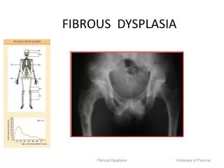
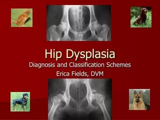
Jeff Binder 2011. Skeletal Dysplasia. Achondroplasia. Squared off pelvis Champaign glass pelvis (caused by anterior pelvic tilt) Squared scapula Widest pedicle spacing at L1 tapering to smallest spacing at L5 (reverse from normal)

E N D
Jeff Binder 2011 Skeletal Dysplasia
Achondroplasia • Squared off pelvis • Champaign glass pelvis (caused by anterior pelvic tilt) • Squared scapula • Widest pedicle spacing at L1 tapering to smallest spacing at L5 (reverse from normal) • Rhizomelia: “short root”, Single long bones (humerus, femur) are short but normal diameter • Bullet nose vertebra: rounded front of vertebrae by defective development of growth plate • Scalloped posterior vertebra: caused by duralectasia • Canal stenosis: 3rd decade • DJD: 3rd decade • Trident hand: separation of 3rd and 4th fingers • Hyperlordotic lumbar spine • Small foramen magnum: MOST LETHAL ASPECT
Osteopetrosis • “BONE WITHIN A BONE” • Fetal bone (primordial) never retreats • “chalk bone” appearance (very dense on film) • Erlenmeyer flask deformity: flared metaphysis of long bones • Dental caries and abscess formation associated • Sandwich vertebra (also called rugger jersey) • Anemia is the most lethal aspect • Reduction in red marrow due to decreased medullary bone • Increased urine Ca levels • Normal bone scans
Melorheostosis • Normal Cortical bone distributed improperly • Strong bones • Bone grown inside and outside of normal bones • Only dysplasia that does this • Dripping wax appearance • No anemia • Cortical thickening and wavy appearance • Can cause peripheral nerve entrapment
OsteogenesisImperfecta • Mucopolysaccharidoses • Brittle bone disease • NOT sclerosing • Arachnodactyly: Long finger/toe appearance due to thinner bones • Brachydactyly: short finger/toe appearance due to premature growth plate closure • Sheppard’s crook deformity: hook like deformity of the femure • Blue sclera: caused by brown choroid visualized through abnormal • Dental problems • Bluish gray to yellowish brown opalescent teeth • Malformed dentin leading to easy chipping and severe caries • Osteoporosis • Wormian bones: not pathognomonic of OI • Multiple fractures: causes excess callus formation on bones • Biconcave vertebral bodies • Undertubularization: narrow bones
Osteopoikilosis • Spotty bone disease • Benign • Asymptomatic • Normal cortical bone growth • Completely within bony margins • If not within bony margins look for another diagnosis • R/O osteoblastic mets in patients over 50yrs • Bone scan will be normal in osteopoikilosis; abnormal in osteoblastic mets (hot spots) • MRI will show a void in signal • Cortical bone is black on MRI therefore areas of cortical bone growth are black
Spondyloepiphyseal Dysplasia • “heaped up vertebra” or “humped shaped vertebra” • Caused by the non-ossification of the ring epiphysis • Thin disk spaces • Short vertebral bodies= short stature; Platyspondly • Premature DJD • Hip and knee invovlement • Subchondralchondrosis
Progressive Diaphyseal Dysplasia • Engelmann’s Disease • Cortical thickening of long tubular bones • CORTICAL BONE ADDED ONLY TO THE INTERIOR OF BONES • Poor muscular development • Increased osteoblastic activity • Bone scan will show • Fusiform widening of only the Diaphysis
Pyknodysostosis • Sclerosing and brittle bone disease • Altered craniofacial ratio • “elfin like features”; small face, normal sized head • Obtuse angle of the jaw • Acroosteolysis: reabsorption of the distal phalanges • Madelung’s deformity • Bowing and overgrowth of the radius contributing to abnormal radioulnar articulations • Dental carries with possible osteomyelitis of mandible
Marfan’s Syndrome • Arachnodactyly: elongation of the fingers without an increase in width • Lens (eye) dislocation • Aortic aneurysms • 45% acquire scoliosis • Tall thin people • Positive thumb sign • Pectisexcavatum: sternum close to the spine “caved in” • 1/3 have congenital heart disease • Atrialseptal defect is the most frequent • Failure to produce normal collagen • Poor quality • Dental carries
CleidocranialDysostosis • Midline structure deformities • Clavicularhypoplasia • Pubic symphysis midline deformity • Spina bifida • Cleft palate • Wormian bones • Widening of coronal and saggital sutures “hot cross bun appearance” • Funnel shaped chest • Hypoplastic and tapered distal phalanges • Biconvex vertebral bodies
Other presentations • Brachycephaly: “short skull” • Premature coronal suture closure • P-A distance decreased • Scaphocephaly: “boat head” • Premature saggital suture closure • S-I distance decreased • Synphalagism • No joints in a finger; finger 1 continuous bone • Syndactyly: “mitten hand” • No soft tissue separation in the hand
Trichorhinophalangeal • Hair, nose, finger • Course hair • Long nose • Finger abnormalities
Stippled epiphysis • Fragmented primary ossification center • Benign