Download
1 / 19
240 likes | 614 Views
Picornaviridae. Foot and mouth disease – oral mucus discharge Genera – major difference between the various genera is the stability at low pH Apthovirus – unstable below pH 7 Rhinovirus – unstable at pH 5 and below

E N D
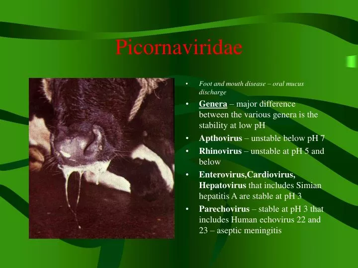
Picornaviridae • Foot and mouth disease – oral mucus discharge • Genera – major difference between the various genera is the stability at low pH • Apthovirus – unstable below pH 7 • Rhinovirus – unstable at pH 5 and below • Enterovirus,Cardiovirus, Hepatovirus that includes Simian hepatitis A are stable at pH 3 • Parechovirus – stable at pH 3 that includes Human echovirus 22 and 23 – aseptic meningitis
Picornaviridae • Stability – important in the epidemiology of the disease • VERY STABLE • If protected by mucus or feces, and shielded from direct sunlight, they are relatively heat stable at normal temps • Apthoviruses and rhinoviruses are less stable, but high humidity can sustain droplets for several hours • Apthoviruses – • Food and mouth disease – Ruminants and swine • Equine rhinovirus 1 – horses – systemic dz.
Picornaviridae • Enterovirus – • Bovine enteroviruses 1-7 bovine subclinical infections • Porcine enterovirus 1 swine polioencephalomyelitis • Porcine enteroviruses 2-11 Swine SMEDI, diarrhea, pericarditis • Swine vesicular disease Swine Swine vesicular disease • Avian enteroviruses Chicken Avian encephalomyelitis • Avian enteroviruses Ducks and turkeys Hepatitis • Rhinovirus- • Bovine rhinovirus 1-3 Cattle Mild rhinitis • Human rhinovirus >100 Humans Common Cold • Cardiovirus - • Encephalomyocarditis swine etc. Encephalomyocarditis • Unclassified virus - • Equine rhinovirus 2 Horses Rhinitis
Foot and Mouth Disease – Apthous fever, Epidemic aphthae • Foot and mouth disease – oral lesions • Aphtha – Gr. Small ulcer • Distribution – currently, North and Central America, Oceania, and the Caribbean – DISEASE FREE • Endemic areas – • Asia, Africa, Middle East and most of South America • Hosts – cloven hooved domestic and wild animals • Cattle, sheep, goats, swine, buffalo • NOT HORSES
Foot and Mouth Disease • Lesions of the hard palate and gingiva • Etiologic agent – Aphthovirus – 7 serotypes A,O,C,SAT-1, SAT-2 and SAT-3 (S.African territories), and ASIA-1 and 80 subtypes • NO CROSS PROTECTION AMONG SEROTYPES • Virus has been shown to survive for 14 days in a stall and for as long as 20 weeks on sacks and hay • Virus in muscle is inactivated in 48 hours of slaughter but survives for longer periods in bone marrow, viscera, blood clots
Foot and Mouth Disease • Foot and mouth disease – lesions of the hock region • Infectivity is destroyed by high temps and UV light • Virus is sensitive to acid and alkaline pH, stable at pH between 6-9, basic disinfectants work well • NaOH, Na carbonate, citric acid, acetic acid • Transmission – inhalation, infected animals generate large volumes of aerosols • Direct contact between infected and susceptible animals by contaminated animal products – meat, milk, semen etc.
Foot and Mouth Disease • Foot and mouth – sloughing of the hoof epidermis • Transmission cont. - • Garbage with uncooked meat and bones from infected animals has been a high source of infection in pigs • Virus can be spread over long distances, country to country, by asymptomatic carriers or animals incubating the disease – virus shedding begins 24 hours prior to observance of clinical signs • Birds, rodents and arthropods have been implicated in the spread of the disease
Pathogenesis – Foot and Mouth • After inhalation – virus replicates in pharynx, viremic spread to the organs follows, REPLICATION OCCURS IN THE EPITHELIAL CELLS OF MUCOSA and skin, causing characteristic lesions • Initial hyperemic areas develop into vesicles, some of which may coalesce to form large blisters. The vesicles are filled with clear, yellow fluid • Myocardial lesions – small gray foci and streaks of irregular size and shape in the myocardium, giving the MYOCARDIUM A STRIPED APPEARANCE, the so-called TIGER HEART are the most common cause of FATAL FMD in young calves, lambs, goats, pigs and buffaloes. • Carrier state – can persist in the pharynx for up to 2 years in cattle and up to 6 months in sheep. There is no carrier state in swine.
Clinical Features – Foot and Mouth Disease – Cattle • Clinical Features – Most severe in cattle and swine ; Sheep and goats develop subclinical infections • Cattle – • IP – 2-8 days; Course 2-3 weeks. • Morbidity high, Mortality less than 5% • Fever, anorexia and depression within 24 hours • Infection causes profuse salivation, lip smacking and are lame • Vesicles are present on the tongue, lips, gums and palate, teats, rumen pillars, coronary band, and interdigital areas. • Ulceration leads to secondary bacterial infection • Abortion – pregnant animals and mastitis with about 25% drop in milk production
Clinical Features – Swine and Ruminants • Swine Clinical Features • Aerosols contain high levels of virus • Vesicles and erosions of snout, lips, and tongue, coronary band • Sheep, goats, and other wild ruminants Clinical Features • Mostly asymptomatic • Disease is usually mild and is characterized by foot lesions and lameness • Diagnosis – • Differentials – vesicular stomatitis, swine vesicular disease, vesicular exanthema, bluetongue, rinderpest, bovine viral diarrhea-mucosal disease • Virus isolation – Vesicular fluid, blood, esophageal and pharyngeal fluid • Cell cultures of bovine origin • ELISA – vesicular fluid or tissues; antibody detection • Virus identification – Virus neutralization, AGID, and complement fixation test
Foot and Mouth Disease Immunity, Control • Cattle recovering from FMD are usually immune to reinfection with the same virus type for a year or longer, but immunity is NOT lifelong • Immunity – type specific, recovered animals can be infected immediately with any of the other serotypes and develop disease • Control – FMD is notifiable disease • Endemic countries – • Vaccination using inactivated and modified live virus vaccines. Quarantine if outbreak • Disease free countries – • Quarantine and slaughter; disposal of carcasses- burning/deep burial,decontamination of premises • Federal law prohibits the importation of animals or products from infected countries unless they are cooked or dried • Zoonosis – Most infections subclinical – fever, anorexia, vesicular lesions on skin and/or mucous membranes followed by recovery
Porcine Polioencephalomyelitis Teschen/Talfan Disease • Severe devastating, neurologic disease of pigs in the Teschen district of Czech Republic • Milder form, Talfan disease is more common and occurs worldwide • Hosts – Swine of all ages are susceptible, however, clinical disease is usually limited to pigs less than 12 weeks of age • Etiologic agent – Porcine enterovirus 1 – Variation in virulence among different strains • Transmission – Ingestion of virus • Virus can survive for months in the environment
Teschen/Talfan Disease • Pathogenesis – • Following ingestion, virus replicates in the GIT and associated lymphoid tissues • There is NO DESTRUCTION OF GUT EPITHELIUM, however, virus is shed into the feces for several weeks • In pigs infected with virulent strains, viremia occurs and results in spread of infection to the CNS • Clinical Features – • IP – 1-4 weeks, course 2 weeks • Tremors, incoordination beginning with hindlimbs, paralysis, prostration, convulsions, coma, death • Mortality may reach 75% • Mild form – Ataxia associated with hind limb paresis followed by recovery
Teschen/Talfan Disease - Vaccine • Diagnosis – • Virus isolation. Brain. Inoculation into porcine cell cultures • FAT staining of infected cell culture for rapid diagnosis • Identification of virus using virus neutralization assay • Pathology – NO SIGNIFICANT GROSS LESIONS ARE OBSERVED AT NECROPSY • Paired serum samples – 4 fold increase in antibody titer • Control – • inactivated and attenuated vaccines are available commercially • Vaccination not practiced in the US • Quarantine and hygiene used primarily
Porcine Enteroviruses 2-11 • Porcine Enteroviruses 2-11 • Frequently isolated from the feces of NORMAL swine and also from sine with diarrhea and pericarditis • There have been isolated from cases of stillbirth, mummification, embryonic death and infertility – SMEDI
Avian Encephalomyelitis “Epidemic Tremor” • Worldwide • Mostly chickens 1-3 weeks of age • Mild encephalomyelitis is seen in turkey poults, quail and pheasants • Etiologic agent – Avian enterovirus • One serotype. Strains may vary in virulence • Transmission – mainly fecal-oral route • Through eggs during a short viremic phase – 1 week – in infected breeder hens – no disease in adults
Avian Encephalomyelitis – Avian enterovirus • Clinical Features – • Main signs are unsteadiness, sitting on hocks, tremors especially of the head and neck, prostration, blindness, paralysis, coma and death • Recovered birds may have CNS deficiencies – may have to be destroyed • Mortality rate may exceed 50% • Diagnosis – viral antigen immunofluorescent stain • Virus isolation – brain. Yolk sac of embryonated eggs/ cell culture • Pathology – No gross lesions seen at necropsy • Control – Either depopulation or vaccination • An attenuated live-virus vaccine administered in the drinking water to breeding hens ensures passively immune progeny. • Maternal antibodies protect chicks for the first few weeks of life
Encephalomyocarditis – rodent spread • Etiologic agent – • Cardiovirus. One serotype. • RODENTS ARE THE NATURAL HOSTS OF THE VIRUS. • The virus infects humans, swine, monkeys, cattle and horses • MOST SEVERE IN YOUNG PIGLETS • Transmission – • Swine are infected by eating rodent urine and feces • Clinical features – • Subclinical in weaned pigs and adults • Sudden death – myocardial failure, encephalomyelitis in young pigs • Anorexia, depression, difficulty breathing • Mortality approaches 100% in very young pigs • In utero – fetal death
Encephalomyocarditis • Diagnosis – • Pathognomonic cardiac lesions – myocardial infarcts esp. in the right ventricle • FAT staining of the affected tissues • Virus isolation – heart and brain • Prevention and Control – • Rodent control. • Outbreaks usually run short courses with immunity occurring in surviving animals • An inactivated vaccine aids in the prevention of reproductive failures in swine