Download
1 / 94
940 likes | 962 Views
Explore how cells respond to signals, from receiving them through receptors to transducing responses. Discover communication mechanisms in multicellular organisms. Investigate oxytocin's impact on bonding in prairie voles and its potential effect on human behavior.

E N D
7 Cell Communication and Multicellularity • 7.1 What Are Signals, and How Do Cells Respond to Them? • 7.2 How Do Signal Receptors Initiate a Cellular Response? • 7.3 How Is the Response to a Signal Transduced through the Cell? • 7.4 How Do Cells Change in Response to Signals? • 7.5 How Do Cells in a Multicellular Organism Communicate Directly?
7 Cell Communication and Multicellularity • In prairie voles, oxytocin and vasopressin released by brain cells bind to other brain cells and trigger life-long bonding and extensive care of offspring. Opening Question: Does oxytocin affect caring behavior in humans?
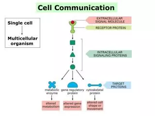
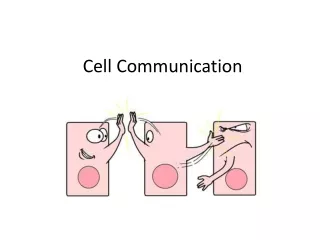
7.1 What Are Signals, and How Do Cells Respond to Them? • All cells process information from the environment. • The information can be a chemical or a physical stimulus, such as light. • Signals can come from outside the organism or from neighboring cells.
7.1 What Are Signals, and How Do Cells Respond to Them? • To respond to a signal, a cell must have a specific receptor that can detect it. • A signal transduction pathway is the sequence of events that lead to a cell’s response to a signal.
7.1 What Are Signals, and How Do Cells Respond to Them? • In large multicellular organisms, signals reach target cells by diffusion or by circulation through the blood. • Chemical signals often in low concentrations.
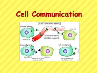
7.1 What Are Signals, and How Do Cells Respond to Them? • Autocrine signals affect the cells that made them. • Juxtacrine signals affect only adjacent cells. • Paracrine signals affect nearby cells. • Hormones travel to distant cells, usually via the circulatory system.
7.1 What Are Signals, and How Do Cells Respond to Them? • A signal transduction pathway involves a signal, a receptor, and responses. • Only cells with the necessary receptors can respond to a signal. • Responses may involve enzymes and transcription factors that are activated or inactivated to bring about the response.
7.1 What Are Signals, and How Do Cells Respond to Them? • Crosstalk: signal transduction pathways can be interrelated. • Pathways can branch; one activated protein may activate multiple pathways. • Multiple pathways can converge on a single transcription factor. • One pathway may be activated while another is inhibited.
7.2 How Do Signal Receptors Initiate a Cellular Response? • Receptor proteins have very specific binding sites for chemical signal molecules, or ligands. • Binding the ligand causes the receptor protein to change shape. • The binding is reversible and the ligand is not altered.
7.2 How Do Signal Receptors Initiate a Cellular Response? • Receptors (R) bind to their ligands (L) according to the law of mass action: R + L ↔ RL • Binding and dissociation (de-binding) have rate constants (K1 and K2).
7.2 How Do Signal Receptors Initiate a Cellular Response? • A rate constant relates the rate of a reaction to the concentration of the reactants: Rate of binding = K1[R][L] Rate of dissociation = K2[R][L]
7.2 How Do Signal Receptors Initiate a Cellular Response? • At equilibrium, the rate of binding equals the rate of dissociation: K1[R][L] = K2[R][L] • or
7.2 How Do Signal Receptors Initiate a Cellular Response? • KD is the dissociation constant, a measure of the affinity of the receptor for its ligand. • The lower the KD, the higher the binding ability of the ligand and receptor. • Very low KD values allow receptors to bind their ligands at very low ligand concentrations.
7.2 How Do Signal Receptors Initiate a Cellular Response? • Many drugs function as ligands. • The KD value of a drug’s binding can be taken into consideration when determining dosage levels.
7.2 How Do Signal Receptors Initiate a Cellular Response? • Inhibitors (or antagonists) can also bind to receptor proteins. • Caffeine is similar to adenosine and binds to the same receptors. Adenosine initiates a signal transduction pathway in nerve cells that reduces brain activity. • Caffeine “ties up” the adenosine receptors, allowing continued nerve cell activity.
7.2 How Do Signal Receptors Initiate a Cellular Response? • Receptors can be located in the cytoplasm or in the cell membrane. • Intracellular receptors: Small or nonpolar ligands can diffuse across the cell membrane (e.g., estrogen). • Membrane receptors: Large or polar ligands bind to cell membrane receptors (e.g., insulin).
7.2 How Do Signal Receptors Initiate a Cellular Response? • Types of plasma membrane receptors in eukaryotes: • Ion channels • Protein kinase receptors • G protein-linked receptors
7.2 How Do Signal Receptors Initiate a Cellular Response? • Ion channel receptors: Channel proteins that allow ions to enter or leave a cell. • Signals can be chemical ligands such as hormones, sensory stimuli such as light, or electric charge differences. • The acetylcholine receptor on muscle cells is a gated ion channel.
7.2 How Do Signal Receptors Initiate a Cellular Response? • Protein kinase receptors: • Some receptors become protein kinases—they catalyze phosphorylation of themselves and/or other proteins. • The insulin receptor phosphorylates itself and other insulin response substrates, which initiates insertion of glucose transporters into the plasma membrane.
7.2 How Do Signal Receptors Initiate a Cellular Response? • G protein-linked receptors: Ligand binding changes the shape of the cytoplasmic region, which binds to a G protein. • G proteins: Mobile membrane proteins with three subunits. They bind GDP (guanosine diphosphate) and GTP (guanosine triphosphate).
7.2 How Do Signal Receptors Initiate a Cellular Response? • GTP-subunit separates from G protein and moves through plasma membrane until it encounters an effector protein. • Binding activates the effector, which causes a change in cell function. • GTP is hydrolyzed to GDP.
7.2 How Do Signal Receptors Initiate a Cellular Response? • Intracellular receptors respond to physical signals such as light or chemicals that can cross the cell membrane. • Many are transcription factors. After binding their ligands, they move to the nucleus, bind to DNA, and alter gene expression.
7.3 How Is the Response to a Signal Transduced through the Cell? • Signals sometimes initiate a chain or cascade of events. • Thus, the initial signal can be amplified and distributed and result in several different responses.
7.3 How Is the Response to a Signal Transduced through the Cell? • Protein kinase receptors bind signals called growth factors that stimulate cell division. • One signal transduction pathway was worked out using bladder cancer cells.
7.3 How Is the Response to a Signal Transduced through the Cell? • Bladder cancer cells have an abnormal form of a G protein called Ras. The protein is permanently bound to GTP, causing continuous cell division. • If the abnormal Ras is inhibited, the cells stop dividing. • This led to the development of Ras inhibitors for cancer treatment.
7.3 How Is the Response to a Signal Transduced through the Cell? • Other cancers have abnormalities in other parts of signal transduction pathways. • By comparing defective and normal cells, complete signaling pathways have been worked out.
7.3 How Is the Response to a Signal Transduced through the Cell? • A protein kinase cascade is a pathway in which one protein kinase activates the next, and so on.
7.3 How Is the Response to a Signal Transduced through the Cell? • In a protein kinase cascade: • The signal is amplified at each step. • Information that arrived at the plasma membrane is communicated to the nucleus. • Multiple steps provide specificity. • Different target proteins provide variation in the response.
7.3 How Is the Response to a Signal Transduced through the Cell? • Some signal transduction pathways include small non-protein molecules called second messengers that mediate some steps. • They were discovered in research on the liver enzyme glycogen phosphorylase, which is activated by epinephrine.
Working with Data 7.1: The Discovery of a Second Messenger • Experiments showed that glycogen phosphorylase could only be activated by epinephrine when the entire contents of liver cells were present. • Hypothesis: a cytoplasmic messenger must transmit the message from the epinephrine receptor at the membrane to glycogen phosphorylase in the cytoplasm.
Working with Data 7.1: The Discovery of a Second Messenger • Glycogen phosphorylase activity was measured in liver cell fractions, with or without incubation with epinephrine.
Working with Data 7.1: The Discovery of a Second Messenger • Question 1: • As part of Sutherland’s research, the activity of glycogen phosphorylase was measured in various liver cell fractions, with or without incubation with epinephrine. The table shows the results. Explain how these data support the hypothesis that there is a soluble second messenger that activates the enzyme.
Working with Data 7.1: The Discovery of a Second Messenger • Question 2: • Propose an experiment to test whether the factor (second messenger) that activates the enzyme is stable on heating (and therefore probably not a protein), and give predicted data.
Working with Data 7.1: The Discovery of a Second Messenger • Question 3: • The second messenger, cAMP, was purified from the hormone-treated membrane fraction. Propose experiments to show that cAMP could replace the membrane fraction and hormone treatment in the activation of glycogen phosphorylase, and create a table to show possible results.
7.3 How Is the Response to a Signal Transduced through the Cell? • These experiments confirmed a second messenger—cyclic AMP (cAMP). • cAMP is produced from ATP by the enzyme adenylyl cyclase. • Adenylyl cyclase is activated via a G protein-linked epinephrine receptor.