Download

1 / 37
370 likes | 502 Views
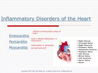
DR. NAILA ALI Assistant Professor OPHTHALMOLOGY. CATEGORIES, COMMON INFECTIVE AND INFLAMMATORY DISORDERS. Categories. Congenital Anomalies Inflammations Disorders of position. Trauma Tumours. Inflammations/ Infections. Stye Chalazion Lid cellulitis Preseptal orbital cellulitis

E N D
DR. NAILA ALI Assistant Professor OPHTHALMOLOGY CATEGORIES, COMMON INFECTIVE AND INFLAMMATORY DISORDERS
Categories. • Congenital Anomalies • Inflammations • Disorders of position. • Trauma • Tumours
Inflammations/ Infections • Stye • Chalazion • Lid cellulitis • Preseptal orbital cellulitis • Blephritis Anterior Posterior
Treatment Options for Trichiasis 1. Epilation - but recurrences within few weeks 2. Electrolysis - but frequently repeated treatments required 3. Cryotherapy - for many lashes 4. Laser ablation - for few scattered lashes 5. Surgery - for localized crop resistant to other methods
Stye (Common Boil) • Small abscess, staph infection of the eyelash, gland of Zeis or Moll • Tender, inflammed swelling in lid margin, single or multiple, may involve entire lid margin– preseptal cellulitis • Treat with hot compresses, removal of eyelash or systemic antibiotics in severe cases • Drain if puss points
Chalazion (Tarsal cyst ormebomian cyst) • Chronic inflammatory granuloma of mebomian gland—blockage and accumulation of secretions • One or more glands involved, mainly children and young adults involved • Swelling, heaviness, irritation • blurring if large- induced astigmatism
Chalazion (Tarsal cyst or mebomian cyst) • Small, cystic, hard swelling a little away from the lid margin, fixed to tarsus, non-tender • No signs of inflammation,no lymphadanopathy • Small may resolve, may remain the same, • may burst on skin- fistula • may infect- internal hordeolum. • treatment • Surgery • steroid injections • Leave alone the small ones.
Signs of chalazion (meibomian cyst) Painless, roundish, firm lesion within tarsal plate May rupture through conjunctiva and cause granuloma
Histology of chalazion Epithelioid Multinucleated cells giant cells Multiple, round spaces previously containing fat with surrounding granulomatous inflammation
Injection of local anaesthetic Treatment of chalazion Incision & curettage Insertion of clamp
Acute hordeola Internal hordeolum ( acute chalazion ) External hordeolum (stye) • Staph. abscess of • Meibomian glands • Staph. abscess of lash follicle • and gland of Zeis or Moll • Tender swelling at lid margin • Tender swelling • May discharge through skin • May discharge through skin • or conjunctiva
Lid cellulitis • Etiology • Multiple styes • Insect bites • Trauma Clinical features whole of the lid is involved tender induration may lead to abscess formation/ skin necrosis Treatment Systemic antibiotics Drainage
CHRONIC MARGINAL BLEPHARITIS 1. Anterior • Staphylococcal • Seborrhoeic 2. Posterior • Meibomianitis • Meibomian seborrhoea
Blephritis • Usually chronic infection of the lid margin • Common external eye disease • Causes—not clear but staph infection and sebhorrea play a part • Associated with tear film instability • Anterior and posterior variety Anterior blephritis—staph or ulcerative and Sebhorroeic or squamous
Staph Anterior Blephritis • Chronic infection of the bases of the lashes resulting in tiny intrafollicular abscesses • Secondary dermal and epidermal ulceration and tissue distruction • More common in children but may affect any age group • Females more affected than males • Unhygienic conditions and dietary factors involved
Staph Anterior Blephritis—Cont. • Symptoms: Sourness of lid margin Lacrimation, itching and photophobia • Signs: Yellow crusts at the roots of the lids On removing the crusts, small ulcers may appear Fall of the lashes—either not replaced or abnormal replacement
Complications If not treat may lead to Poliosis, madarosis, trichiasis, tylosis • Treatment: General: Improvement in general health Balanced diet Correction of refractive errors Local: Broad spectrum antibiotic ointment, steroid ointment Artificial tears. Treat the sequlae.
Staphylococcal blepharitis • Chronic irritation worse in mornings • Hyperaemia and telangiectasia of anterior • lid margin Scales around base of lashes (collarettes) • Scarring and hypertrophy if longstanding
Sebhorroeic Ant Blephritis • Disorder of the glands of Zeis and Moll • Sebhorreoa may involve scalp, eyebrows, nasolabial folds, retroauricular area and sternum. • Oily type and dry type (true seborrheoa)
Sebhorroeic Ant Blephritis • Symptoms: less severe, discomfort in the eyes, lacrimation, tear film instability (stinging) • Signs: Shiny, waxy appearance of the anterior lid margin, dandruff like desquamation lid epidermis (yellow, greasy scales) • Treatment: Lid hygiene— artificial tears Scalp treatment with antidandruff lotion
Seborrhoeic blepharitis • Shiny anterior lid margin • Greasy scales • Hyperaemia of lid margin • Lashes stuck together
Posterior Blephritis • May present as: *Dysfunction of the mebomian glands. *Isolated mebomian seborrhoea and primary mebomenitis. *Combination of ant. Seb. Blephritis and meibominitis
Primary Mebominitis • Diffuse inflammation around glands • Associated with acne rosacea (2/3rd) or Seb. Dermatitis (1/3rd) Signs: *Pouting of gland orifices with dome of secretions—may become solid. (tooth-past like) *Obliteration of ducts—dilatation (thick, round, vascularised, notched Posterior border) *Secondary changes include papillary conjunctivitis, punctate epitheliopathy, and tear film instability (stinging)
Meibomianitis Meibomian cyst formation Thickened posterior lid margin
Mebomian Seborrheoa • Dilated mebomian glands—easily expressed • Tear film is oily and foamy • In severe cases—mebomian foam at medial canthus.
Meibomian seborrhoea Oily and foamy tear film Oil globules over meibomian gland orifices
Treatment: A: Systemic antibiotics Tetracycline, Doxocycline, Erythromycin B: Others: Lid hygiene, topical steroids, artificial tears. C: Local measures: Warm compresses, mechanical expressions of secretions
Complications trichiasis, madarosis , poliosis Recurrent styes Tear film instability Marginal keratitis
Trauma • Lid margin • Lid tissue • Loss of lid tissue
Eyelid haematoma Usually innocuous but exclude associated trauma to globe or orbit Orbital roof fracture if associated with subconjunctival haemorrhage without visible posterior limit Basal skull fracture - bilateral ring haematomas (‘panda eyes’)