Download
1 / 59
660 likes | 1.14k Views
INVERSION OF ANTENATAL PYRAMID. Dr.Malathi G Prasad Dr. Bavaharan Fetal Medicine Centre Trichy. Pregnancy Care. 19 th century –pregnancy care for wealthy 20 th century – High MMR & IMR prenatal care 16 , 24 , 28 & fortnightly

E N D
INVERSION OF ANTENATAL PYRAMID Dr.Malathi G Prasad Dr. Bavaharan Fetal Medicine Centre Trichy
Pregnancy Care • 19th century –pregnancy care for wealthy • 20th century – High MMR & IMR prenatal care • 16 , 24 , 28 & fortnightly • No rationale on timing or clinical content
AIM • Early screening of Fetal Aneupliodies • Early Diagnosis of Fetal Abnormalities • Early Screening for Miscarriage and Stillbirth • Early Screening for Preeclampsia • SGA • Preterm Delivery
New 1st Trimester Scan Protocol • NT • Nasal Bone • Early survey • Doppler • DuctusVenosus • Tricuspid Valve flow • Hepatic artery flow • Uterine artery Doppler
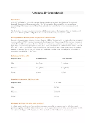
Timing of Ultrasound and Biochemistry • first-trimester combined screening for trisomy 21 is to perform biochemical (B-HCG ,PAPP-A) ultrasonographic testing & include maternal age One-stop clinic for assement of Risk – OSCAR The ideal gestation for OSCAR is 12 weeks because the aim of the first-trimester scan is not just to screen for trisomy 21 but also to diagnose an increasing number of fetal malformations
Assessment • If Combined Risk 1:50 Invasive testing • <1:1000 – Screen Negative • 1:50 to 1:1000 • absence of the nasal bone, • increased impedance to flow in the ductusvenosus and tricuspid regurgitation
Biochemical • In trisomy 21 , Serum free β-hCG is about twice as high and PAPP-A is reduced to half compared with euploid pregnancies • Trisomies 18 and 13,Serum free β-hCGand PAPP-A are decreased • In cases of sex chromosomal anomalies, maternal serum free β-hCG is normal and PAPP-A is low • Triploidies -- free β-hCG and PAPP-A is low
Protocol for measurement of nuchal translucency • 11 to 13 weeks 6days. • CRL45 and 84mm. • The magnification of the image should be such that the fetal head and thorax occupy the whole screen. • A mid-sagittal view of the face • The fetus should be in a neutral position, with the head in line with the spine • Distinguish between fetal skin and amnion. • The widest part of translucency must always be measured. • Measurements should be taken with the inner border of the horizontal line of the callipers placed ON the line that defines the nuchal translucency thickness - the crossbar of the calliper should be such that it is hardly visible as it merges with the white line of the border, not in the nuchal fluid. • Turn the gain down. • A new approach for the measurement of NT which improves the accuracy of measurements, is with the use of a semi-automated technique.
Risk of a cardiac defect • NT measurement Cardiac risk <95th centile 0.16% 2.5-3.4 mm 1% 3.5-4.5 mm 3% 4.5-5.4 mm 7% 5.5-6.4 mm 20% Above 6.5 mm 30%
Fetal anomalies associated with increased NT • Major cardiac defect • Diaphragmatic hernia • Exomphalos • Body stalk anomaly • Skeletal defects. • Genetic syndromes • Congential adrenal hyperplasia • Fetal akinesia sequence • Noonan syndrome • Smith-Lemli-Opitz • Spinal muscular atrophy
Interpretation of NT Increased NT Points to a possible Abnormality UNIVERSAL SCREENING – Combined Test -- OSCAR
Protocol for assessment of the fetal nasal bone • The gestational period must be 11 to 13 weeks and six days. • The magnification • A mid-sagittal view of the face should be obtained. • The ultrasound transducer should be held parallel to the direction of the nose and should be gently tilted from side to side to ensure that the nasal bone is seen separate from the nasal skin. • The echogenicity of the nasal bone should be greater that the skin overlying it. In this respect, the correct view of the nasal bone should demonstrate three distinct lines: the first two lines, which are proximal to the forehead, are horizontal and parallel to each other, resembling an "equal sign". The top line represents the skin and bottom one, which is thicker and more echogenic than the overlying skin, represents the nasal bone. A third line, almost in continuity with the skin, but at a higher level, represents the tip of the nose. • When the nasal bone line appears as a thin line, less echogenic than the overlying skin, it suggests that the nasal bone is not yet ossified, and it is therefore classified as being absent.
If other parameters are suspicious – Invasive testing Interpreting Nasal Bone
Open Spina Bifida In almost all cases of open spina bifida associated Arnold-Chiari malformation Consequence of leakage of cerebrospinal fluid into the amniotic cavity and hypotension in the subarachnoid spaces caudal displacement of the brain and obstructive hydrocephalus. In the second trimester of pregnancy, the manifestations of the Arnold- Chiari malformation are the lemon and banana signs Caudal displacement of the brain is apparent at 11–13 weeks in the same midsagittal view of the fetal face the lower part of the fetal brain between the sphenoid bone anteriorly and the occipital bone posteriorly can be divided into the brain stem in the front and a combination of the fourth ventricle and cistern magna in the back In fetuses with open spina bifida, the brain stem diameter is increased and the diameter of the fourth ventricle-cisterna magna complex is decreased. Hypoplasia of vermis & cerebellum can also be picked up in 1st trimester Intracranial Translucency
1ST Trimester Detectable Abnormalities • Body stalk anomaly, anencephaly, alobarholoprosencephaly, exomphalos, gastroschisismegacystis. • Amelia or Phocomelia
1st trimester Undetectable abnormalities • because they are manifested only during the second or third trimester of pregnancy, • microcephaly, • agenesis of the corpus callosum, • semilobarholoprosencephaly, • hypoplasia of the cerebellum or vermis, • cystic adenomatoidmalformation or • pulmonary sequestration • bowel obstruction.
Protocol for the assessment of the ductusvenosus • The gestational period must be 11 to 13 weeks and six days. • The examination should be undertaken during fetal quiescence. • The magnification of the image fetal thorax and abdomen occupy the whole image. • A right ventral mid-sagittal view of the fetal trunk should be obtained and color flow mapping should be undertaken to demonstrate the umbilical vein, ductusvenosus and fetal heart. • The pulsed Doppler sample volume should be small (0.5-1.0 mm) • The insonationanglelessthan 30 degrees. • The filter should be set at a low frequency (50-70 Hz) • The sweep speed should be high (2-3 cm/s) so that the waveforms are spread allowing better assessment of the a-wave.
Anomaly & fetal Echo Interpretation of Ductus
DUCTUS VENOSUS FLOW NO OF CASES REVERSAL ANEUPLOIDIES CARDIAC ANOMALIES 942 18 4 9
Anomaly & fetal Echo Interpretation of Tricuspid regurgitation
Uterine artery PI • Aim of identifying women at high-risk for subsequent development of preeclampsia • screening by maternal history may detect only about 30% of those that will develop preeclampsia for a false positive rate of 5% • Improve pregnancy outcome because intensive maternal and fetal monitoring in such patients would lead to an earlier diagnosis of the clinical signs of the disease and the associated fetal growth restriction • avoid the development of serious complications through such interventions as the administration of antihypertensive medication and early delivery
Protocol for first-trimester measurement of the uterine artery PI • 11 weeks and 13 weeks and six days. • Sagittal section of the uterus • Identify the cervical canal and internal cervical os • the transducer must be gently tilted from side to side and then colour flow mapping should be used to identify each uterine artery along the side of the cervix and uterus at the level of the internal os. • Pulsed wave Doppler • sampling gate set at 2 mm to cover the whole vessel • ensuring that the angle of insonation is less than 30º.
Interpretation of Uterine artery UA PI – INCREASE – impaired trophoblastic invasion of maternal spiral arterioles & their conversion to wide non muscular channels depend on Maternal Vasomotor tone
Statistics Prospective study at FMC – 18 months Total Sample -- 942 Screen Positive – 109 Invasive tests done for 91 Positive aneuploidies – 13
Early Screening for Miscarriage and Stillbirth • increased fetal NT thickness, • reversed a-wave in the fetal ductusvenosus and • low maternal serum PAPP-A PREVENTION No intervention for miscarriage Still Birth-closer monitoring
Early Screening for Preeclampsia • 2% pregnancies Evolving evidence • degree of impaired placentation and • Incidence of adverse fetal and maternal short-term and long-term consequences of preeclampsia are inversely related to the gestational age
PRE ECLAMPSIA • Maternal characteristics • Maternal Weight • Obesity • Preexisting HT or DM • Mean arterial pressure • Uterine artery PI markers
Preecl …. contd • Biochemical tests • PAAP-A , PGF , inhibin A Sensitivity – 90% PREVENTION Close Surveillance Aspirin
GDM • Maternal Characteristics • Risk increses with maternal age & BMI • Family history GDM • Biochemical markers • Adiponectin • Sex hormone binding globulin • GCT AT 11-13 – Cut off -130mg/dl • Sensitivity—75% Dietary advice , drugs