Download

1 / 32
350 likes | 582 Views
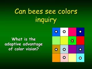
An Optical Illusion Apart from the green and white background, how many colors can you see?. Illusions. Slipped Disk. The Brain. parietal lobe. Occipital Lobe. frontal lobe. Temporal Lobe.

E N D
An Optical Illusion Apart from the green and white background, how many colors can you see?
parietal lobe Occipital Lobe frontal lobe Temporal Lobe
1. Frontal pole 2. Superior frontal sulcus 3. Middle frontal gyrus 4. Superior frontal gyrus 5. Precentral sulcus 6. Longitudinal cerebral fissure 7. Precentral gyrus 8. Postcentral gyrus 9. Central sulcus 10. Postcentral sulcus 11. Occipital pole
1. Frontal pole of left cerebral hemisphere 2. Olfactory bulb 3. Olfactory tract 4. Orbital gyri and sulci 5. Straight gyrus 6. Temporal pole of left cerebral hemisphere 7. Olfactory trigone 8. Optic nerve 9. Optic chiasma 10. Anterior (rostral) perforated substance 11. Optic tract 12. Tuber cinereum with infundibulum 13. Oculomotor nerve 14. Mamillary body 15. Uncus of parahippocampal gyrus 16. Basis pedunculi 17. Basilar sulcus of pons 18. Trigeminal nerve 19. Abducens nerve 20. Pyramid of medulla oblongata 21. Facial nerve 22. Vestibulocochlear nerve 23. Glossopharyngeal nerve 24. Vagus nerve 25. Cranial roots of accessory nerve 26. Spinal roots of accessory nerve 27. Rootlets of hypoglossal nerve 28. Flocculus 29. Ventral rootlets of 1st cervical spinal nerve 30. Pyramidal
1. Short gyri of insula 2. Central sulcus of insula 3. Circular sulcus of insula 4. Long gyrus of insula
1. Frontal forceps 2. Corpus callosum commissural fibers 3. Short arcuate fibers 4. Occipital forceps 5. Indusium griseum 6. Medial longitudinal stria 7. Lateral longitudinal stria Corpus callosum, its radiation, and indusium griseum displayed from above
Olfactory senses • "The VNO appears to be a much more primitive structure that uses a different set of molecular machinery than the main olfactory system ,"
Brain Scans PET scan on the left shows two areas of the brain (red and yellow) that become particularly active when volunteers read words on a video screen: the primary visual cortex and an additional part of the visual system, both in the back of the left hemisphere. Other brain regions become especially active when subjects hear words through ear-phones, as seen in the PET scan on the right.
How you Hear!!!!! • How it Works:The auditory canal can resonate and amplify sounds within a frequency range of about 2000 Hz to 5500 Hz by up to a factor of 10. • Successive compressions and rarefactions of air reaching the eardrum result in a change in pressure between the outer ear and the middle ear. The Eustachian tube helps to keep the middle ear at atmospheric pressure. • The difference in pressure between the sound wave Sound waves travel through the meatus (auditory canal) to the tympanic membrane (ear drum striking the outer surface of the eardrum and normal atmospheric pressure on the inside of the eardrum causes the eardrum to vibrate. • Within the middle ear, vibrations travel through three small bones (the hammer, anvil, and stirrup) to the cochlea. The bones act as interlocking levers which amplify the force of the eardrum striking the hammer. The oval window of the cochlea is smaller than the eardrum. This causes a further amplification of the sound vibration, upt 20 times at some frequencies. • The semicircular canals act as miniature accelerometers. They also help to maintain a sense of balance by responding to gravity and changes in acceleration. The hair-like structures (dendrites) in the cochlea resonate at various different frequencies. The vibrations stimulate neurons to produce electrical impulses which are sent along the auditory nerve to the brain for processing.
C A B
Brain Scans PET scan on the left shows two areas of the brain (red and yellow) that become particularly active when volunteers read words on a video screen: the primary visual cortex and an additional part of the visual system, both in the back of the left hemisphere. Other brain regions become especially active when subjects hear words through ear-phones, as seen in the PET scan on the right.
How We See!!!! Light rays reflected by an object—for example, a pencil—enter the eye and pass through its lens. The lens projects an inverted image of the pencil onto the retina at the back of the eye. Signals produced by rod and cone cells in the retina then start on their way into the brain through the optic nerve and reach a major relay station, the LGN (lateral geniculate nucleus). Signals about particular elements of the pencil then travel to selected areas of the primary visual cortex, or V1, which curves around a deep fissure at the back of the brain. From there, signals fan out to "higher" areas of cortex that process more global aspects of the pencil such as its shape, color, or motion. Surprisingly, light rays must penetrate two layers of neurons in the retina before reaching the precious rods and cones at the back: a middle layer of bipolar cells, and a front layer of ganglion cells whose long axons (fibers that transmit electrical impulses to other neurons) form the optic nerve leading into the brain.
Safety Tip • Why did the potato where safety goggles?
Even Potatoes have To protect their”EYES”!