Download
1 / 27
370 likes | 780 Views
Nerve Impulses. Chapter 48. Membrane Potential. Is the difference in electrical charge across the cell membrane . Outside of cell is positively charged due to excess of Na+ ions Cells are bathed in a salt solution thus the Na+ and Cl - ions outside of cell

E N D
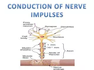
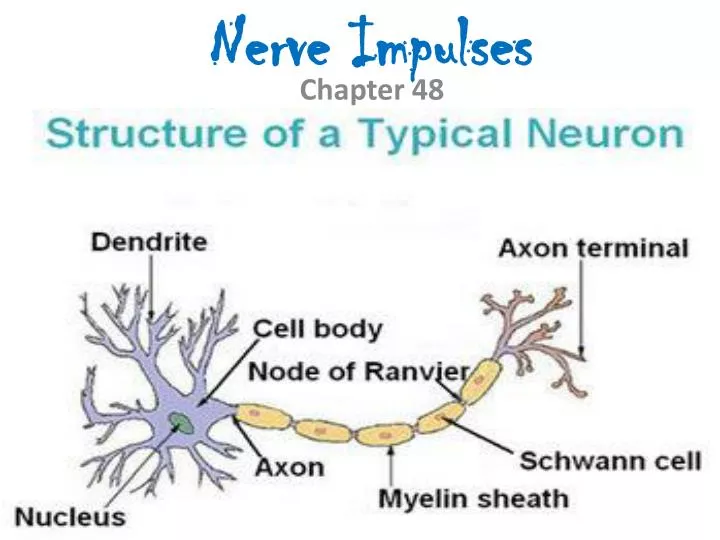
Nerve Impulses Chapter 48
Membrane Potential • Is the difference in electrical charge across the cell membrane. • Outside of cell is positively charged due to excess of Na+ ions • Cells are bathed in a salt solution thus the Na+ and Cl- ions outside of cell • Therefore, inside of cell is negative charged with <K+ to Na+ ions [ ] • Are produced by the movement of ions across the cell membrane • Movement of ions depends on: • the ability of the ions to diffuse through the cell membrane • the concentration of ions inside and outside the cell • the electrical charge of the ions • Is expressed as voltage in mV
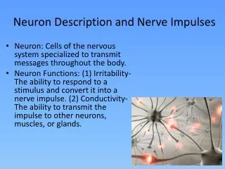
Resting Potential • A neuron at rest is not receiving or sending a signal. • Concentration of negatively charged proteins and positively K+ ions is greater inside the cell than outside. • The [ ] of Na+ ions is greater outside the cell than inside. • [ ] difference between K+ and Na+ ions is due to the sodium-potassium pump which pumps 3 Na+ ions out of the cell to every 2 K+ ions into the cell • Na+ doesn’t readily diffuse through the membrane therefore they accumulate outside the cell. • Negatively charged proteins are too large to diffuse • K+ pass freely through the membrane & tend to diffuse out of the cell • [ ] of K+ inside cell is 140mM and 5mM outside cell thus they move out of cell • Exit of K+ out of the cell and retention of negatively charged proteinsinside the cell cause the interior of the neuron to become negatively charge with respect to the outside. • This difference in charge now is referred to as the resting potentialof the membrane. • Most neurons, the resting potential is - 70mV
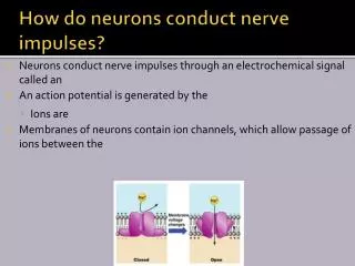
Ions move through membranes by passing through proteins that act as ion channels. • Each ion channel is specific to the ion that passes through it • Certain channels are voltage gated which open or close dependent on the membrane potential • Voltage-gated potassium channels contain positively charged 'paddles‘ that move within the membrane to open the channel. • As ions move into or out of the neuron, they then affect membrane potential
K+ ions continue to diffuse out of the neuron until chemical and electrical forces balance out. • This is accomplished by the negative forces from the Ca- ions and the negative proteins within “pulling” the K+ ions back into the cell • Creates anEquilibrium potential for K+ of -92 mV • Inside of membrane is - 92 mV more negative than the outside
Na+ ion [ ] is 15 mV inside the cell and 150mV outside of the cell • Some Na+ ions can move across open Na+ ion channels into the neuron • This movement makes inside of the neuron a little less negative • Equilibrium potential of Na+ is +62mV
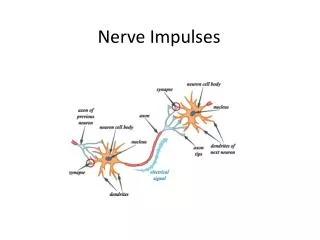
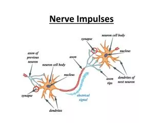
Action Potential • When a dendrite of a neuron is stimulated, the permeability of the neuron’s membrane changes • Membrane becomes permeable to Na+ ions which rush into the cell opens voltage-gated channels (only at the nodes of Ranvier) in the membrane which allows Na+ ions to flow into the neuron • Now the interior becomes more positively charged than the outside. • Reversal of polarity across the membrane begins an action potential • Action potential begins where the cell body meets the axon • Action potentials are electrical signals that move along a neuron’s membrane • Animation
Na+ Na+ Na+ Na+ Na+ K+ K+ K+ K+
As the first part of the axon become more positively charged, the change in voltage opens up the next segment of the axon membrane • More Na+ ions enter, moving in a + direction, open up more channels. • + charges are moving down like dominos falling • Axon potentials usually travel only away from the cell and end at the axon terminal • The voltage gates for Na+ close and then the voltage gates for K+ open. • Threshold potential is now reached at -55 mV • Interior again becomes + with respect to outside • Since K+ gates are still open, there is an overshoot of K+ ions out which makes interior even more negative. • Negatively charged proteins draw K+ ions back in due to polarity of charges. • Resting potential is now restored • Neuron can not generate another action potential until the resting potential is restored.This time period is the refractory period
Following the action potential, the [ ] of Na+ is higher inside than when the cell was at rest and the [ ] of K+ ions inside the cell is lower. • Ion channels & the sodium-potassium pump will reestablish the resting concentration of Na+ and K+ ions. • 3Na+ ions will be moved out and 2K+ ions will be moved in. • Neuron will be back to normal -70mV resting potential • To maintain normal resting potential, a continuous supply of ATP will be needed
Salatory Conduction • In myelinated axons, the depolarizing current during an action potential at one node of Ranvierspreads along the interior of the axon to the next node • This enables the next ion-gated channel to open in the next node of Ranvier
Jumping the gap • Neurons are not physically connected to each other • Gap is known as the synaptic cleft • Secretion of neurotransmitters (stored in synaptic vesicles) from the presynaptic neuron will bridge this cleft • Arrival of action potential depolarizes the membrane, opens voltage-gated channels and allows Ca+ ions to diffuse into the terminal • Rise in Ca+ [ ] in terminal causes synaptic vesicles to fuse with membrane, releasing neurotransmitters • Neurotransmitters within the axon terminal fuse with the presynaptic membrane and cross thesynaptic gap/cleft. • Neurotransmitters bind to receptor proteins in the postsynaptic cell’s membranes.
Interaction of neurotransmitter and receptor molecules changes the permeability of the postsynaptic membrane’s ion channels. • Na+ ion channels of postsynaptic membrane causes the membrane potential to become more positive. • If change in membrane potential is great enough, a new action potential is generated in the receiving neuron • Electrical signal continues – Excitatory • If too few Na+ channels open in postsynaptic membrane or allow negative ion channels to open, then membrane potential of receiving neuron will remain negative and no action potential will be generated. • Electrical signal is terminated – Inhibitory
Neurotransmitters get cleared from synaptic clefts shortly after release • Presynaptic neurons reabsorb neurotransmitters to be used again • This reabsorption ensures that the neurotransmitters effects on postsynaptic cells is not continuous
Neurotransmitters • 100 neurotransmitters • Acetylcholine – Excitatory effect • Muscle stimulation • Memory formation • learning • Amino Acids • Glutamate • Excitatory effect for long term memory • Gamma-aminobutryric acid – GABA • Inhibitory effect • Valium binds to GABA receptors • Biogenic Amines • Norepinephrine– excitatory orinhibitory • Dopamine and serotonin – affect sleep, mood, attention and learning • Endophins –Inhibitory • Natural pain blockers • Depress respiration • Produce euphoria Animation