Download
1 / 4
50 likes | 202 Views
BIO 210 Lab Handout #8 – Articulations & Body Movements

E N D
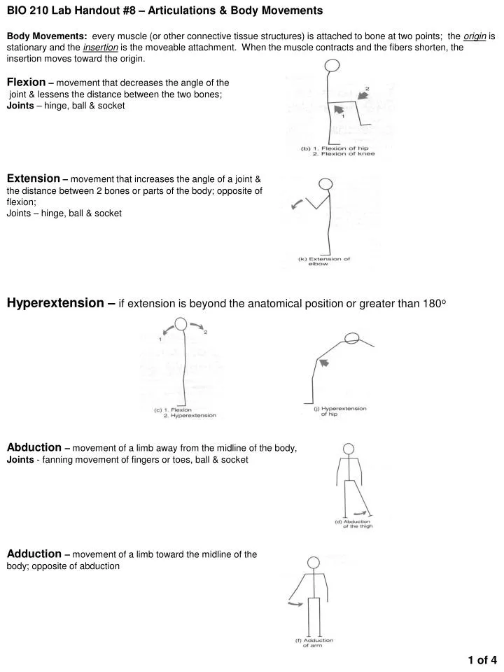
BIO 210 Lab Handout #8 – Articulations & Body Movements Body Movements: every muscle (or other connective tissue structures) is attached to bone at two points; the origin is stationary and the insertion is the moveable attachment. When the muscle contracts and the fibers shorten, the insertion moves toward the origin. Flexion – movement that decreases the angle of the joint & lessens the distance between the two bones; Joints – hinge, ball & socket Extension – movement that increases the angle of a joint & the distance between 2 bones or parts of the body; opposite of flexion; Joints – hinge, ball & socket Hyperextension – if extension is beyond the anatomical position or greater than 180o Abduction – movement of a limb away from the midline of the body, Joints - fanning movement of fingers or toes, ball & socket Adduction – movement of a limb toward the midline of the body; opposite of abduction 1 of 4
Rotation – movement of a bone around its longitudinal axis without lateral or medial displacement; Joints – Atlanto-axial, ball & socket; Circumduction – combination of flexion, extension, abduction & adduction; proximal end of the limb remains stationary while the distal end moves in a circle Joints – ball & socket Movements of the Hand Pronation – movement of the palm of the hand from an upward-facing position to a down-ward facing position; anterior to posterior Supination: - movement of the palm from a posterior position to an anterior position; opposite of pronation Movements of the Foot Dorsiflexion – movement of the ankle joint in a dorsal direction (standing on one’s heels) Plantar Flexion - movement of the ankle joint in which the foot is flexed downward (standing on one’s toes; pointing the toes) Inversion – movement that results in the medial turning of the sole of the foot Eversion – movement that results in the lateral turning of the sole of the foot: opposite of inversion 2 of 4
Articulations Suture – irregular edges of the bones interlock & are united by very short connective tissue fibers 1. Frontal 2. Sagittal 3. Lamboidal 4. Squamous Symphyses – bones are connected by a broad, flat disc of fibrocartilage 1. Symphysis pubis 2. Sacroiliac joint 3. Intervertebral joints Synovial – joints in which the articulating bone ends are separated by a joint cavity containing synovial fluid; all are characterized by the following structures 1. Joint surfaces enclosed in a sleeve of fibrous connective tissue (articular capsule) 2. Synovial membrane produces synovial fluid for lubrication; reduce friction 3. Articulating surfaces of the bones are covered with hyaline cartilage 4. Capsule is reinforced with ligaments 5. Fibrocartilage pads may be present within the capsule Femur Lateral condyle Medial condyle Sacroiliac joint Patella Fibula Tibia Anterior cruciate ligament Olecranon process Pubic symphysis Humerus Radius Trochlear notch Ulna Ulna Trochlea Hinge – rounded process of one bone fits into the concave surface of another to allow movement in one plane, usually flexion and extension 1. Tibiofemoral (knee)- between the femoral condyles and the C-shaped minisci (semilunar cartilages) of the tibia Medial & lateral condyles of femur Anterior cruciate ligament – attaches to the anterior intercondylar tubercle of the tibia; passes posteriorly, laterally, and upward to attach to the femur on the medial side of the lateral condyle 2. Humeroulnar (Elbow) – close gripping of the trochlea by the ulna’s trochlear notch that forms the hinge a) Olecranon process of the ulna b) Trochleaof the humerus 3 of 4
Ball-and-socket – ball shaped head of one bone fits into a cuplike depression of another; allows movement in all directions and pivotal rotation 1. Coxal Joint (Hip) – formed by the articulation of the spherical head of the femur with the deeply cupped acetabulum of the Os Coxae (hip bone) a) Acetabulum of os coxae b) Headof femur Os coxa Acetabulum Head 2. Glenohumeral Joint (Shoulder) – large hemispherical head of humerus fits in the shallow glenoid fossa of the scapula like a golf ball sitting on a tee a. Glenoid fossaof scapula b. Headof humerus Head Scapula Glenoid fossa Humerus Femur 4 of 4