Download

1 / 30
300 likes | 459 Views
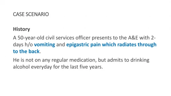
A CASE SCENARIO. by. Dr.Syed Hunain Riaz P.G M2. Slide 001. HISTORY. Case History. A 14 years old boy of very low socioeconomic background presented with 1. Shortness of breath - ------- About 10 years 2. Generalized weakness ----- About the same duration. Slide 002.

E N D
A CASE SCENARIO by Dr.Syed Hunain Riaz P.G M2 Slide 001
Case History • A 14 years old boy of very low socioeconomic background presented with 1. Shortness of breath--------About 10 years 2. Generalized weakness-----About the sameduration Slide 002
History of presenting illness The boy was was in his usual state of health when he was about 4 years old when he gradually developed shortness of breath. It was exertional in nature. • He did not have orthopnea or PND. • There is h/o palpitations associated with the dyspnea. • No h/o chest discomfort, body swellings or hemoptysis. • No h/o syncopal events. • H/o yellowish discoloration of eyes for the last many years, no h/o abdominal distention, no h/o unconsciousness • He started to develop generalized weakness and as such was unable to perform simple activities.
No h/o blood in stools, no h/o alternating bowel habits or worms in stool. PAST HISTORY • Non/diabetic, non/diabetic, non-asthmatic. Has had blood transfusions three times over period of 10 years. FAMILY HISTORY • No h/o diabetes, IHD, asthma or TB in the family, no h/o any disease in his siblings. Has 3 brothers, all older than him, all healthy.
DRUG HISTORY No h/o any drug intake and no allergies to any medicine • SOCIOECONOMIC HISTORY Belongs to a lower class family who earn their bread by farming
General Physical exam • A young boy of average built with prominent facial bony features ( slightly protuding forhead, prominent zygomatic area ) well oriented, sitting comfortably in bed. • Vitals and Exam findings : BP: 90/50 mmHg Pulse: 90/min R/R: 18/min Temp: Afebrile Jaundice: ++(lime colored) Pallor: +++ Cyanosis: - Clubbing: -
Lymph nodes: - JVP: Not Raised Pedal edema: - Palmar erythema: - Neck veins: Not distended Koilonychia: - • 2 shallow ulcers(2-3 cm each) with clean margins and yellowish discharge on dorsum of right foot.
Systemic Exam Cardiovascular: • Apex beat in left fifth ICS, normal in character • Soft S1 and S2, soft A2 and loud P2 • Pansystolic murmur heard in mitral area, radiating to axilla. • Pansystolic murmer heard in tricuspid area, increasing in intensity with inspiration
Gastrointestinal: • Abdomen soft, non tender, no pulsations or veins visible • Upper border of liver in right 5th ICS, lower edge palpated about 4 fingers below right hypochondrium, extending into the epigastrium, non-tender, and smooth surfaced. Liver span about 18 cm • Spleen palpated 2 fingers below left hypochondrium towards the umbilicus, non tender and smooth surfaced. • No free fluid on in abdomen on examination
CNS: • Grossly intact Respiratory: • Normal chest shape and movements • Chest expansion normal • NVB with no added sounds Muskoloskeletal: • NAD
Differential Diagnosis based on History\Exam • Anemia ( Iron deficiency? Hemolytic? ) • Lymphoproliferative disorder? • Valvular heart disease ( leading to long standing anemia )?
CBC with peripheral smear • Hb: 3.4 g/dl • Rbc: 1.3 ×10^12/l • Hematocrit: 8% • MCV: 75 fl • MCH: 24 pg N ( 26-32 ) • MCHC: 31 g/dl N ( 30-35 ) • Platelets: 265×10^9/l • WBC: 7×10^9/l • Neutrophils, monocytes, eosinophils, basophils and lymphocytes all within normal limits • Retic count: 1%
RBC morphology: Hypochromia: +++ Microcytosis: ++ Anisocytosis: +++ Poikilocytosis: +++ Target cells: ++ ESR:150 mm/1st hour LDH: 550 IU ( upto 450 ) URINE C/E: • Normal pH, no sugars, no proteins. • No hemoglobinuria, no hemosiderin pigments. • Urobilinogen normal, bilirubin not detected • No blood cells.
Liver function tests: • Bilirubin total: 2.1 mg/dl • Bilirubin direct: 1.3 ( 0.1-0.7 ) • Bilirubin indirect: 0.8 ( 0.1-0.4 ) • ALT: 56 IU • AST: 41 IU • ALK phosphatase: 394 IU/l ( 100-270 ) • Proteins: 5.4 g/dl • Albumin: 2.7 • Globulin: 2.7 • PT: 16/13 • aPTT: 36/33 Renal function tests: Urea: 17mg/dl Creatinine: 0.5 mg/dl
Stool exam: No ova or parasites, negative for occult blood. USG ABDOMEN\ PELVIS: • 18.5 cm Liver with normal echotexture, intra/extrahepatic biliary channels normal, no focal lesion • Spleen 16 cm ( enlarged ) • KUB normal • No fluid or lymph nodes seen in peritoneal cavity CHEST XRAY: Enlarged heart, lungs and costophrenic angles clear ECG: Normal axis and rhythm, symmetric T-wave inversions in anterior chest leads
Echocardiography: • Mild dilatation of cardiac chambers • Slightly increased pulmonary arterial pressure • Normal valves but functional MR and TR present • Normal biventricular function
Based on clinical and investigation data, there was a high suspicion of a long standing hemolytic anemia (extravascular hemolysis), specifically “ THALASSEMMIA”, so a hemoglobin electrophoresis was ordered…..
Hemoglobin Electrophoresis: • HbA: 53.4 % • HbF: 44.2 % • HbA2: 2.4 %
CONCLUSION The final diagnosis was: ß-THALASSEMIA INTERMEDIA/ MAJOR
Thalassemias • A heterogenous group of inherited disorders of hemoglobin synthesis resulting decreased or absent hemoglobin chains synthesis • Classified into alpha and beta thalassemias. • Varied pattern of inheritance • Decreased synthesis of ß-chains result in over production of alpha chains, and also gamma and delta chains leading to the finding of fetal and HbA2 type of hemoglobin in ß-thalassemias
Treatment • Thalassemia major\ intermedia patients are treated on the following lines: • BLOOD TRANSFUSIONS • CHELATION THERAPY ( DESFERIOXAMINE ) • ALLOGENIC BONE MARROW TRANSPLANTATION Splenectomy may be needed if BMT is not feasible for reducing the RBC destruction in the spleen