Download
1 / 25
260 likes | 518 Views
Síndrome Antifosfolípido y embarazo. Anticuerpos Anti-Fosfolípidos. La primera evidencia de los aFL fue en 1952, con los falsos positivos para la detección de Sífilis. Antígeno: cardiolipina-colesterol-leutina.

E N D
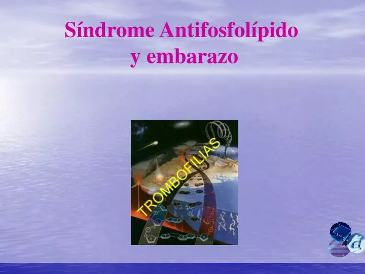
Síndrome Antifosfolípido y embarazo
Anticuerpos Anti-Fosfolípidos • La primera evidencia de los aFL fue en 1952, con los falsos positivos para la detección de Sífilis. • Antígeno: cardiolipina-colesterol-leutina. • En 1998 en un panel Internacional en Sapporo (Japón), se estableció los criterios para definir el Síndrome Antifosfolípido (SAF). • El SAF presenta trombosis arterial y/o venosa, abortos a repetición y trombocitopenia, al menos en dos determinaciones, con un intervalo superior a 8 semanas. • Entre los antígenos que se distinguen para los aFL se encuentran: ß2GPI, protrombina, anexina, proteína C y S entre otras.
Anexina • La pérdida recurrentes de embarazos es un problema frecuente en la práctica obstétrica. • Hoy casi cerca del 50% de los casos no se han aclarado, mientras que la otra mitad se le atribuye a: • Alteraciones genéticas • Diabetes • Infecciones materno Fetal • Los aFL pueden desencadenar: • Preclampsia severa • Retardo en el crecimiento • Parto prematuro • Trombosis y abortos espontáneos
Técnicas para la detección de los Anticuerpos Anti-Fosfolípidos • ELISA en fase sólida • Anticoagulante lúpico (AL) • Tiempo de Protrombina diluido (dTP) • Tiempo de coagulación con kaolín (KCT) • Tiempo de veneno de víbora Rusell (dRVVT)
Anexina(Cont) • Forman una familia de proteínas que tienen afinidad por fosfolípidos y fue reconocida en 1990. • Existen cerca de 100 tipos diferentes de anexinas y solo 13 en mamíferos. Figura 1 • Su estructura esta compuesta por secuencias repetitivas consistente en 70 aminoácidos. • Anexina II une plasminógeno y activador del plasminógeno, activando el proceso de la fibrinolisis. • Las atleraciones adquiridas o congenitas de las anexinas reciben el nombre de “Anexinopatías”.
Anexina(Cont) • La anexina mas estudiada es la V, la cual posee una actividad anticoagulante, por desplazamiento de las proteínas de la coagulación. • Se une a los fosfolípidos con ayuda de calcio, además, se une a heparan sulfato. • En la unión de la anexina V a la membrana un residuo de triptofano del domio 3, penetra en ella. • Participa en la formación del fagosoma, inhibe Fosfolipasas citosolicas y proteínas kinasas. • La membrana apical del sincitiotrofoblasto expresa FS, donde se uniria la anexina V. Figura 2
Schematic illustration of the fetal-maternal interface in humans and mice. The placenta, representing the main interface between the mother and fetus, is composed of two parts: the trophoblast of embryonic origin, and the decidua of maternal origin. • In the human placenta, the syncytiotrophoblast cover of the villi is the main site for all maternofetal transfer and secretory functions, and some of the extravillous cytotrophoblast migrate to an endovascular location, where they can form a new vessel lining, in spiral arteries in particular. • Red arrows indicate the blood flow to and from the placenta via maternal arteries or veins, respectively. V: villous trophoblast; IVS: intervillous space; CMA: mouse central maternal artery; S : spongiotrophoblast; L: labyrinthine trophoblast; UC: umbilical cord; MD : maternal decidua.
Efectos de los aFL en la Anexina V • Tanto los aFL y la Anexina V tienen afinidad por los fosfolípidos aniónicos. • En el SAF los aFL desplazarían a la anexina V de la superficie fosfolípidica con lo que se desencadenaría los mecanismos trombogénicos. Figura 3
Figure 3. B, Annexin-V, in the absence of aPL antibodies, serves as a potent anticoagulant by forming a crystal lattice over the anionic phospholipid surface, shielding it from availability for assembly of the phospholipid-dependent coagulation complexes. C, In the absence of annexin-V, aPL antibody-ß2GPI complexes can prolong the coagulation times, compared with control antibodies. This occurs via antibody recognition of domains I or II on the ß2GPI, which results in dimers and pentamers of antibody-ß2GPI complexes having high affinity for phospholipid via domain V. These high-affinity complexes reduce the access of coagulation factors to anionic phospholipids. This may result in a lupus anticoagulant effect in conditions where there are limiting quantities of anionic phospholipids. D, In the presence of annexin-V, aPL antibodies, either directly or via interaction with protein-phospholipid cofactors, disrupt the ability of annexin-V to form ordered crystals on the phospholipid surface. This results in a net increase of the amount of anionic phospholipid available for promoting coagulation reactions. The aPL-cofactor complexes expose significantly more phospholipids by disrupting the annexin-V shield than they block by direct binding. This manifests in the net acceleration of coagulation in vitro and in thrombophilia in vivo.
b2 Glicoproteína I • En 1990 se reconoció que los aFL estaban dirigidos contra otras proteínas como la b2 GPI. Figura 4 • Peso molecular de 50 kDa. • Es altamente glicosilada y con un alto contenido de prolina. • Concentración plasmática es de 200 ug/ml y en un 40% se encuentra asociada a lipoproteínas. • Posee cinco dominios repetidos, de los cuales el 5° tiene una región carboxiterminal, es altamente catiónico por su gran contenido de lisina y arginina. Figura 5 • Además son críticos los residuos de cisteina en las posiciones 281 y 288. • La sustitución de estas moléculas de cisteina o del quinto dominio por ingeniería genética anula la unión de la b2GP-1 a fosfolípidos aniónicos.
b2 Glicoproteína I (cont.) • Se une a sustancias con carga negativa como fosfolípidos, heparina, lipoproteínas, y plaquetas activadas. • La b2GPI es un anticoagulante natural ya que inhibe la vía intrínseca de la coagulación, la actividad de protrombinasa, y la agregación plaquetaria dependiente de ADP. • Estos anticuerpos se asocian más a trombosis venosa. Figura 6 • No existe diferencia con el tipo de inmunoglobulina y el tipo de trombosis con respecto a los anticuerpos anti-cardiolipina. Figura 7
Figure 5. Structure of human blood plasma ß2GPI. A, Ribbon model of ß2GPI based on crystal structure: the protein is composed of an extended chain of 5 SCR domains having a "fishhook" appearance. The structure of SCR domain V deviates from the standard fold of the 4 other domains and forms the putative phospholipid-binding site. ß-Strands are shown in red and helices in green. (Reprinted from Bouma B, de Groot PG, van den Elsen JM, Ravelli RB, Schouten A, Simmelink MJ, Derksen RH, Kroon J, Gros P. Adhesion mechanism of human ß2-glycoprotein I to phospholipids based on its crystal structure. EMBO J. 1999;18[19]:5166–5174, with permission of Oxford University Press). B, The structural data suggest a simple membrane-binding mechanism in which the cationic patch of domain V has an affinity for anionic phospholipid. The stretch of Ser311 to Lys317 forms a hydrophobic loop that inserts into the lipid bilayer and positions Trp316 at the interface region between the acyl chains and the phosphate headgroups of the lipids, thereby anchoring the ß2GPI in the membrane. (Reprinted from Bouma B, de Groot PG, van den Elsen JM, Ravelli RB, Schouten A, Simmelink MJ, Derksen RH, Kroon J, Gros P. Adhesion mechanism of human ß2-glycoprotein I to phospholipids based on its crystal structure. EMBO J. 1999;18[19]:5166–5174, with permission of Oxford University Press).
Figure 6.Anti– b2-glycoprotein I antibodies and thrombosis: OR with 95% CI grouped according to the type of thrombosis. (A) Arterial thrombosis. (B) Venous thrombosis. (C) Any thrombosis. n.s. indicates not significant. 4394 patients and 1973 controls. Analysisin relation to the isotype showed IgG anti–b2-glycoproteinI antibodies were significantly associated with thrombosis in20 (61%) of 33 cases, IgM in 7 (47%) of 15 cases, IgA in 3 (100%)of 3 cases, and the G/A/M isotypes (when no distinction waspossible) in 4 (44%) of 9 cases. There were 10 studiesthat included multivariate analysis: 2 of them confirmedthat IgG anti–b2-glycoprotein I antibodies were independentrisk factors for venous thrombosis.
Anti-Protrombina • En 1959 Loeligeret al, describió un anticuerpo que prolongaba los tiempos de coagulación, porque se unen al complejo protrombina/fosfolípido. Figura 8 • Al absorber el plasma con BaSO4concluyo que la protrombina era uno de sus cofactores. • En 1984 Edson et al, fue el primero en demostrar la existencia de los anticuerpos antiprotrombina, también Flecket al, encontro un 74% en pacientes con Anticoagulante Lupico. • Existen diferentes epitopes en la protrombina. Figura 9 • Presentan una alta asociación con trombosis Figura 10
Fig 9. Different pathways of prothrombin activation. (*) Indicates active site exposure. Inverted "Y" indicates g-carboxyglutamic acid. • Entre los epitopes se encuentran en: • Protrombina • Pretrombina 1 (Carboxilo terminal de la Protrombina) • Fragmento 1 (Segmento amino-terminal de la protrombina) • Fragmento 1.2 (Segmento amino-terminal de la protrombina) • Como reconoce la región amino terminal de los factores Vitamina K dependiente también reconoce a la proteína C y S. • También Plasminógeno.
Figure 10.Antiprothrombin antibodies and thrombosis: OR with 95% CI grouped according to the type of thrombosis. (A) Arterial thrombosis. (B) Venous thrombosis. (C) Any thrombosis. n.s. indicates not significant. 4394 patients and 1973 controls. Overall, 17 (37%) of 46 associations were significant: 3 of11 associations with arterial thrombosis, 7 of 18 with venousthrombosis, and 7 of 17 with any thrombosis reached significance.Analysis in relation to the isotype showed IgG antiprothrombinantibodies were associated with thrombosis in 11 (26%) of 24cases, the M isotype in 2 (14%) of 14 cases, and the G/A/M isotypesin 4 (50%) of 8 cases. There were 8 studiesthat did a multivariate analysis: 2 of them confirmed thatantiprothrombin antibodies were independent risk factors forthrombosis and 3 others showed that they added to the risk borneby lupus anticoagulants or anticardiolipin antibodies.
Trabajos a Futuro • Anticuerpos anti-proteína S, en pacientes con LES • Anticuerpos anti-b2GPI en LES. • Anticuerpos aPT como riesgo de TVP • Anticuerpos aFL en pacientes con Stroke • Anticuerpos anti-b2GPI y fosfatidilserina como predictores de trombosis arterial • Anticuerpos anti-b2GPI y aPT afectan la formación del coágulo • Anticuerpos aFL en abortos recurrentes. • Anticuerpos anti-Fosfatidilserina en mujeres con hijos prematuros • Anticuerpos anti-proteína S, en pacientes Abortos • Anticuerpos anti-anexina V en pacientes con trombosis • Inhibición competitiva de la unión de la anexina a cardiolipina y generación de • trombina en pacientes no seleccionados con trombosis venosa • Polimorfismo C-1T incrementa los niveles de anexina V
Pacientes: No embarazadas (n:50, 30 10 años) • Primer trimestre (10 sem gestación, n:30, 30 6 años) • Segundo trimestre (24 sem, gestación, n: 50, 30 5 años) • Tercer trimestre (33-34 sem, n: 40, 30 v 6 años) • Antes del parto (n:53, 30 5 años) • 3 a 5 días postparto (n: 67, 30 5 años) • Pacientes 146 mujeres con 2 o más perdidas fetales, 99 controles • Pacientes con niños pprematuros, 290 controles • La gran mayoría de las investigaciones usan ELISAs “Home Made”, encontrándose una diferencia en resultados • Al comparar kits comerciales v/s ELISAs “Home Made”, para la detección los anticuerpos Anti-b2 GPI no muestran ventajas. • No existen kits comerciales para la detección de anticuerpos antiprotrombina • Existen al menos 3 ELISAs para detección de anticuerpos: • Protrombina • Protrombina + Fosfatidilserina • Protrombina + Fosfatidilserina + Calcio • Placas g-Irradiadas o Poliestireno