Download
1 / 33
330 likes | 638 Views
Screening for Diabetic Retinopathy This presentation lasts approximately 10 minutes. Diabetic Retinopathy. Diabetic retinopathy is damage to blood vessels and tissue in the retina (the layer at the back of the eye) caused by diabetes.

E N D
Screening for Diabetic Retinopathy This presentation lasts approximately 10 minutes.
Diabetic retinopathy is damage to blood vessels and tissue in the retina (the layer at the back of the eye) caused by diabetes. • Diabetic retinopathy can affect anyone with diabetes, whether they have type 1 or type 2. • The condition can be well advanced before any reduction of vision is experienced.
Diabetic retinopathy is the main cause of blindness in the16 - 65 year old age group. • Early detection and regular monitoring are essential to reduce the risk of progression and improve the chances of successful laser treatment.
Good blood sugar and blood pressure control can reduce the risk of diabetic retinopathy and other complications of diabetes.
The retina is the innermost layer of the eye. • The retina contains photoreceptor cells (rods and cones) these cells work like the film in a camera by recording light which enters through the pupil. • This light information is sent from the retina, along the optic nerve and into the brain where the image is formed.
Main Features of the Retina OPTIC NERVE (OPTIC DISC 1.5mm) FOVEA MACULA
Optic Nerve (Optic Disc) - The nerve which connects the eye to the brain, and brings the retina its main blood supply. • Macula - Responsible for fine detail central vision (reading, writing etc.) and colour vision. • Fovea - The centre of the macula which provides the sharpest point of human vision.
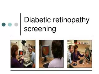
Screening for Diabetic Retinopathy Using a Digital Camera
Regular screening for diabetic retinopathy is the best way to reduce the risk of visual impairment caused by diabetes. • ***** PCT/Hospital provide annual screening for anyone with diabetes registered with a local GP. • The screening service uses a digital fundus camera (camera used to photograph the inside of the eye) which produces instant pictures. These can be shown to the patient during their appointment.
Your Screening Appointment • When you arrive you will have to check in at reception. • You will be asked to take a seat in the waiting area until the nurse or screening assistant calls your name.
First, your vision will be tested using a standard wall chart, the same sort that you use when you visit your optician.
Eye drops are then inserted into your eyes which dilate your pupils and allow a clear view for photography. This may sting at first.
IT IS NOT ADVISABLE TO DRIVE FOR UP TO 4 HOURS AFTER THE EXAMINATION AS THE EYE DROPS CAN CAUSE TEMPORARY BLURRING OF VISION AND SENSITIVITY TO BRIGHT LIGHT! It is advisable to bring sunglasses with you to wear when you leave.
When your pupils are dilated (after about 15 mins) the screener will take 4 photographs, 2 in each eye. The screener will then show you the photographs and point out any signs of diabetic retinopathy. The results will be sent to your GP.
A letter outlining the results of the screening appointment and any further action required will be sent to you within 4 weeks of your appointment.
Micro aneurysms - Are small red spots. These are caused by a swelling of very small capillary vessels in the retina, they are an early sign of diabetic retinopathy. Micro aneurysms should be monitored every 12 months.
superficial flame shaped haemorrhage deep round haemorrhage Small blot haemorrhages Haemorrhages - Are red blots varying in size and shape. These are small bleeds within the retina or near the surface. There are several types . Haemorrhages should be monitored every 3, 6 or 12 months depending on severity.
Some Other Signs of Diabetic Retinopathy Hard exudates - Shiny pale white or yellow sharp edged features. These are fatty deposits caused by leaking fluid. Cotton wool spot - White and fluffy patches. These are scarred nerve fibres near the surface of the retina. Venous loop - A loop in a blood vessel, caused by poor flow of blood. These signs should be assessed by an ophthalmologist.
New blood vessels - These appear wispy and fragile. New blood vessels form as a result of existing vessel damage. These vessels are extremely weak and tend to rupture very easily. This causes scarring and a build up of blood within the eye. New vessels require laser treatment.
Maculopathy - This applies to most of the signs we have already looked at when they occur on the macula, close to the fovea. Hard exudates Haemorrhages Micro aneurysms Early maculopathy requires close monitoring. With hard exudates laser treatment is needed.
Laser treatment uses an intense beam of light directed onto the retina. • The treatment is given by an Ophthalmologist and is given as an outpatient. • There are three main types of laser treatment.
Pan-Retinal Photocoagulation • This is used to treat proliferative retinopathy (lesions which affect most of the retina with the exception of the macula). The laser is pointed at the outer part of the retina to reduce the risk of bleeding and scarring from repeated haemorrhages. This may take several sessions of laser treatment.
Panretinal Photocoagulation (PRP) - applied to treat proliferative diabetic retinopathy.
Focal Laser Treatment • Damaged blood vessels in the macula can leak fluid and exudates (fatty deposits) which damage your central vision. Focal laser treatment (often in combination with grid laser treatment) involves pointing the beam at the leaking blood vessels to prevent further leakage. This leakage and the resulting retention of fluid is known as Macular Oedema.
Grid Laser Treatment • This treatment applies low power laser burns to the retina in a grid pattern. This process removes the fluid from the back of the eye to improve sight by stimulating the cells that normally drain fluid away from the retina.
After laser treatment • After laser treatment you will be followed up by the Ophthalmologist for several months. When the Ophthalmologist is happy that the condition has stabilised you will be returned to the screening service for regular monitoring.
PLEASE CONTACT THE SCREENING SERVICE ON*** **** ****IF YOU HAVE ANY QUERIES OR CONCERNS REGARDING DIABETIC RETINOPATHY. THANK YOU FOR YOUR ATTENTION.