Download
1 / 74
1.02k likes | 2.28k Views
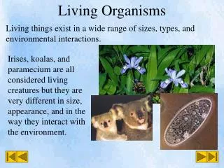
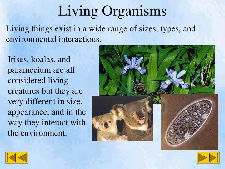
Living Organisms. Living things exist in a wide range of sizes, types, and environmental interactions. . Irises, koalas, and paramecium are all considered living creatures but they are very different in size, appearance, and in the way they interact with the environment.

E N D
Living Organisms Living things exist in a wide range of sizes, types, and environmental interactions. Irises, koalas, and paramecium are all considered living creatures but they are very different in size, appearance, and in the way they interact with the environment.
To classify what is living and what is not living is actually quite difficult. Scientists are still grappling with this argument trying to decide if viruses are indeed living creatures. There are 5 basic characteristics that scientists have agreed on. Living organisms will… • Need energy • Respond to their environment. • Reproduce • Grow • Produce wastes
Every living organism carries out these functions in different ways. They need to use specialized structures to fulfill these functions. Lions use their teeth, mouths, digestive tracks to gain energy. They need to kill another organism to gain their energy. The trees leaves on the other hand collect sunlight and use special chemicals to synthesize energy. Just in the way that they gain energy, lions and trees have very different interactions with their environment.
In our own bodies we have many organs which are specialized to carry out the functions we need to live. Each organ is made from special tissues. These muscle tissues Are made from long bundles on thin striations.
Muscle tissues are common in the body. The skeletal muscles move our limbs. As well, muscle tissues are part of our stomach, heart, and esophagus. The muscle tissues are made from cells. A cell is the basic unit of a living system. Like organs and tissues, each cell is specifically designed to help carry out a function. The muscle cell uses long fibers that relax and contract chemically to complete their function.
The stem, leaves, and roots of this elodea plant are the organs. The stem, leaf, and roots are made from different specialized tissues. These tissues are made from different specialized cells. Plants have very different cells that make up their tissues. The aquatic plant elodea has cells designed to help support the plant as well as make food using sunlight.
Microscopes It is impossible for us to see cells with our eyes. This is why many of our ideas about cells were not discovered until the late 1600’s after the invention of the microscope. A microscope is a device that magnifies objects. Magnification makes objects appear larger. A simple magnifying glass is in a way a type of microscope.
In the late 1600’s, Robert Hooke used a simple one lens microscope to look at a thin slice of oak cork. He saw little rooms that reminded him of the cells where monks lived. Cork cells are dead so he only saw their cell walls.
Around the same time as Hooke, Anton van Leeuwenhoek used simple microscopes to view rain water and blood. He saw in rain water things he called “animalcules” which we now call bacteria. In blood, saw blood cells of different varieties and shapes. Van Leewenhoek made his own simple microscopes grinding lenses to about the size of the head of a pin. They could magnify about 300 x.
German botanist Matthias Schlieden and zoologist Theodore Schwann made many observations of cells in every living tissue they studied from plants, muscles, nerves and many others in the early 1800’s. Schlieden Schwann • Another German scientist, Rudolf Virchow, proposed a cell theory about living things. • All living things are made of cells • Cells are the basic units of structure and function in living things.
All early microscopes used light to view the microscopic world. Mirrors would deflect light through a sample and into the magnifying lens. Microscopes that use light are called light microscopes and are still used today however an electric light source is place beneath the object being studied.
Simple microscopes (with one lens) were the tool of choice for a long time. Compound microscopes (using 2 lenses) had been invented in the 1600’s. The images produced by compound microscopes were blurry due to the relatively poor lens quality. However, a compound microscope can offer greater magnification.
The very best light microscopes can magnify about 2000x. This is not enough to see some of the smaller parts of cells but, the images are in color and single celled organisms can be seen alive. Electron microscopes can magnify today up to 2 000 000x. Electrons are passed and scattered off of objects and then recorded on a photographic plate. The images are black a white and kill the living objects being observed. The first electron microscopes were developed in Germany. The first practical design of an electron microscope was developed in Canada by James Hillier and Albert Prebus.
The eyepiece (ocular lens) is helps magnify the object being observed. It is also what you look though to see your object. The tube holds the eyepiece and objective lenses at the correct distance from each other. As well, it refracts the light through the eyepiece.
The revolving nosepiece holds the objective lenses and allows you to change to different lenses and higher and lower powers of magnification. The arm holds up the revolving nosepiece. It is also what you hang onto when moving a microscope. The objective lenses magnify the object. The low power objective lens is the smallest while the high power lens is the largest.
The stage holds the object you are viewing and moves up and down to focus the object. The stage clips secures the object to the stage so that it doesn’t move around with small bumps and movements of the microscope.
The diaphragm controls the amount of light going through the object and into the objective lens. More light is needed at higher magnifications. The course-adjustmentknob quickly moves the stage to focus the image (usually only used under low power magnification). The fine-adjustment knob slowly moves the stage to focus the image (usually used for higher magnifications).
The light source shines light through the object and into the objective lenses. The base provides a heavy support for the microscope so that small pushes and movements will not juggle the object being viewed. Always have one hand on the base when carrying a microscope.
When you look at an object trough a microscope you see only a small portion of the entire object. What you see through the eyepiece is called the field of view. As the magnification is increased, the amount you see gets smaller.
In order to measure the field of view a ruler needs to be observed under the microscope. An ordinary clear plastic ruler will work. Usually under low power, the millimeter lines on a clear plastic ruler are visible. In this case the low power magnification was 4x and we could see 5.5 mm lines. Therefore, the field of view for the low power is 5.5 mm.
Mag. of low power lens Low power field of view Medium power field of view = Mag. of medium power lens If the low power 4x lens, for a microscope, gives a field of view of 5.5 mm then we can calculate the field of view for the other magnifications. Let’s say the medium power lens has a magnification of 15x. We can use the proportion of magnification to field of view with the following formula. 4x = 5.5 mm Med f. of v. 15x 1.5 mm Med f. of v.
The accuracy of this calculation methods completely depends on how accurate the measurement of the low power field of view. To get more precise calculations of field of view, special and expensive rulers called micrometers are viewed at different magnifications. A micrometer divides a single millimeter into many tiny divisions so accurate measurements can be made.
When we look at microscopic organisms we don’t look at them directly on the lens. Instead, we make a wet mount. First, you place a drop of a sample onto a clear glass slide. The sample contains the objects you want to view. Then you place a clear glass cover slip over the sample. The cover slip will stick to the slide holding down the objects you want to view.
The specimen is sandwiched between the cover slip and the slide. Air bubbles are commonly seen as small round shapes. Being careful when making your wet mount can reduce the appearance of air bubbles. Most cells are transparent. Stains are added to help highlight certain cells and cell parts. This hydra has been stained purple so that it can be easily seen with a microscope. Imperfections in the glass of the slide and lenses are also seen under the microscope.
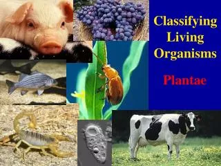
The Cell We have seen the theory that all living tissues are made from cells. This can help us to categorize living things into two broad groups. Unicellular organisms are made from only one cell. There are many different examples that range from animal like carnivores like paramecium and hydra to single celled plants like algae. Many fungi are also unicellular like penicillium.
Organisms made from many cells are multicellular. This group encompasses all other forms of organisms from dust mites to blue whales. We are much more familiar with multicellular organisms due to the fact that we can easily see and interact with them. However, there are far more varieties of single celled bacteria than all other forms of life combined. In fact, the estimate of the total living mass of bacteria far outweigh any other form of life.
Just like animals cells contain structures that complete specific functions. In animals these are called organs. In cells, these are called organelles. Most organelles are invisible to even the best light microscopes Each organelle completes a specific function for the cell. Not all cells have the same organelles however, there are some common characteristics of cell organelles. For example, cheek cells (left) look very different from onion skin cells (right).
The cell membrane separates the interior of the cell and its environment. It also controls the movement of materials in and out of the cell. The membrane is like the cells “skin.” It is visible with a light microscope.
Cytoplasm Cell Membrane Nucleus The cytoplasm is a fluid inside of the cell. It is constantly moving and helps distribute disolved nutrients to different parts of the cell. The cytoplasm is also visible with a light microscope with the use of a colored stain. The cytoplasm is like the cells “blood.”
The nucleus of the cell is usually found at the center. It contains the genetic information in chromosomes. The nuclear membrane is similar to the cellular membrane. Small holes exist on the membrane called nuclear pores. Inside the nucleus is the nucleolus. This small ball produces other organelles called ribosomes. The nucleus is visible with a light microscope and the appropriate stains. It is like the “brain” of the cell because it controls the cells functions.
The endoplasmic rheticulum (ER) is a folded organelle usually near the nucleus. It aids in the transport of materials in the cell. Ribosomes are small organelles that produce long strands of protiens like a teletype. If the ER has ribosomes attached to it we call it the rough ER and if there are no ribosomes we call it the smooth ER. The ER and ribosomes are not usually visible with a light microscope. The ER is like the cells “veins.”
The mitochondrion is a bean shaped organelle with many folds and ridges called cristae. These produce the energy for the cell. Muscle cells would have many mitochondria (plural of mitochondrion) to produce a lot of energy. These are like the “power plants” of the cell.
The Golgi apparatus is a folded organelle that packages materials into balls called vacuoles. Vacuoles can then be transported safely and efficiently throughout the cell. Come vacuoles contain digestive chemicals. These are called lysosomes. A lysosome needs to be kept separate from the rest of the cell otherwise the digestive chemicals would kill the cell. Vacuoles The Golgi apparatus is like the “post office” of the cell. Vacuoles and lysosomes are like the cell’s “stomach.”
The cell wall is only found in plant cells. It is made from cellulose and offers the plant support. It is thick and difficult to transport materials out of the cell wall. Plant cells therefore need large vacuoles to store wastes.
Chloroplasts are only found in plant cells. They are green organelles that convert light energy to chemical energy. The chlorophyll in the chloroplasts in plants is green which is why plants are green.
Cells are cell organelles are very small. Even the largest animals and plants are made from very tiny cells that are close to the same size. Having a small size makes cells very efficient. Firstly, it doesn’t take much energy to transport from the membrane of the cell to the center. Secondly, the surface area to volume ratio is very large if the cell is small. Imagine two cubic cells. One has has a side length of 1mm and the other 10mm.
Fluids and Movement in Cells Cells require materials to exist and complete their necessary functions. They need water, air, food, and a variety of other nutrients. The cell membrane separates the cell from the rest of its environment. It is like our skin. Cell membranes have openings and special passage ways that let materials in and out of the cells. This means that the membrane is selectively permeable. If the membrane didn’t let anything into the cell it would be impermeable.
Fluids and Movement in Cells • An impermeable membrane would be fatal to cells since they couldn’t get their needed nutrients. • A totally permeable membrane would also be fatal since even harmful chemicals could enter and destroy the cell.
One way for a cell to gain and remove materials is by using vacuoles. A vacuole containing waste can approach the plasma membrane and merge with it. An opening is created on the other side and the waste pushed out. The reverse process can also occur where the membrane can grow around and consume some kind of beneficial material.
The process of gradual mixing of particles in fluids is called diffusion. According to the particle theory, since particles of a fluid are in constant motion, when a clump of fluid particles are added to the moving particles of another fluid, they will tend to spread out and intermix. Diffusion is a natural process. When perfume is released in air, it diffuses through the air to eventually spread evenly throughout the room.
The principle of diffusion is that particles will tend to move from areas of high concentration to areas of low concentration. Imagine yourself breathing. When you expel carbon dioxide, the concentration around you is relatively higher than 2 m away from you. Therefore, the carbon dioxide tends to diffuse away from you, which is a good thing otherwise we would simply breath the carbon dioxide back. A cell “breaths” in a similar way. When this amoeba creates carbon dioxide waste the concentration inside the cell is relatively higher than outside the cell. By diffusion the carbon dioxide moves out of the cell.
Cell membranes remain impermeable to most large particles. Water particles, however, are able to flow relatively easily through cell membranes. The diffusion of water across a membrane is called osmosis. When a cell is put into pure water, there is a great tendency for water to rush into the cell (since there is relatively little water inside the cell compared to outside). If this happens too quickly the cell can burst in a process called lysis.
Osmosis can have a significant effect. When there is a relatively large amount of dissolved solute on one side of a membrane the osmotic pressure will push water to even out the concentration of the two sides.
Plants use osmosis and diffusion to gain the nutrients and water they need. If celery is placed in pure water, osmosis pushes water into the stalk. The cell wall prevents lysis and the celery stands upright. If the stalk is place in a salt water solution, then there is relatively more water inside the cells so water rushes out through osmosis. The stalk falls limp. Salty water Pure water
Water and nutrients move through a plant through special tissues called vascular tissues. Phloem tissue moves sugars made by the leaves to the rest of the plant. Xylem tissues move water and dissolved minerals to parts of the plant.
The roots of a plant absorb the necessary water and minerals to make its food. Tiny root hairs have semipermiable membranes that connect to the xylem tissues. Osmotic pressure pushes the water up the xylem to the leaves where most of the photosynthesis (food production) occurs.
The large flat leaves are packed with chloroplasts to gather sunlight and make sugars. Oxygen is allowed to diffuse into the leaf through openings called stomata on the underside of the leaf. Plants need to breath just like us. They use the oxygen in exactly the same way we do to make energy for to do many of the functions they need. This also means they need to expel waste gasses. When the stomata are open, carbon dioxide, oxygen, and water escape. This “breathing out” is called transpiration.