Download
1 / 1
20 likes | 146 Views
Head-to-Head Comparison of Myocardial Perfusion SPECT Imaging and Magnetic Resonance Delayed Enhancement Detection of Myocardial Infarction Marcus Carlsson MD PhD, Håkan Arheden MD PhD Dept of Clinical Physiology, Lund Universtiy Hospital, Sweden

E N D
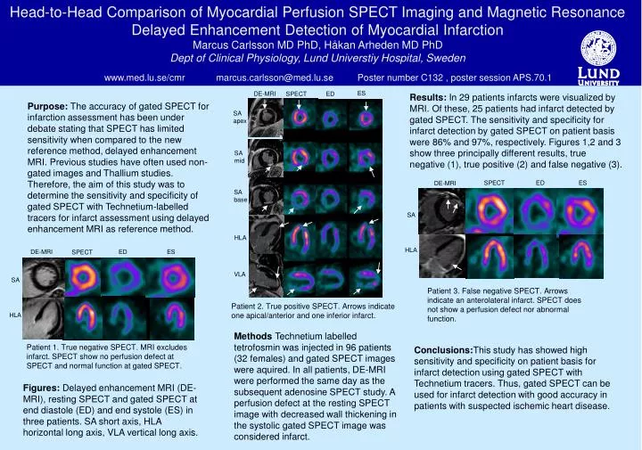
Head-to-Head Comparison of Myocardial Perfusion SPECT Imaging and Magnetic Resonance Delayed Enhancement Detection of Myocardial Infarction Marcus Carlsson MD PhD, Håkan Arheden MD PhD Dept of Clinical Physiology, Lund Universtiy Hospital, Sweden www.med.lu.se/cmr marcus.carlsson@med.lu.se Poster number C132 , poster session APS.70.1 SPECT ED ES DE-MRI ES DE-MRI SPECT ED SA HLA DE-MRI ED ES SPECT Results: In 29 patients infarcts were visualized by MRI. Of these, 25 patients had infarct detected by gated SPECT. The sensitivity and specificity for infarct detection by gated SPECT on patient basis were 86% and 97%, respectively. Figures 1,2 and 3 show three principally different results, true negative (1), true positive (2) and false negative (3). Purpose: The accuracy of gated SPECT for infarction assessment has been under debate stating that SPECT has limited sensitivity when compared to the new reference method, delayed enhancement MRI. Previous studies have often used non-gated images and Thallium studies. Therefore, the aim of this study was to determine the sensitivity and specificity of gated SPECT with Technetium-labelled tracers for infarct assessment using delayed enhancement MRI as reference method. SA apex SA mid SA base HLA VLA SA Patient 3. False negative SPECT. Arrows indicate an anterolateral infarct. SPECT does not show a perfusion defect nor abnormal function. Patient 2. True positive SPECT. Arrows indicate one apical/anterior and one inferior infarct. HLA Methods Technetium labelled tetrofosmin was injected in 96 patients (32 females) and gated SPECT images were aquired. In all patients, DE-MRI were performed the same day as the subsequent adenosine SPECT study. A perfusion defect at the resting SPECT image with decreased wall thickening in the systolic gated SPECT image was considered infarct. Patient 1. True negative SPECT. MRI excludes infarct. SPECT show no perfusion defect at SPECT and normal function at gated SPECT. Conclusions:This study has showed high sensitivity and specificity on patient basis for infarct detection using gated SPECT with Technetium tracers. Thus, gated SPECT can be used for infarct detection with good accuracy in patients with suspected ischemic heart disease. Figures: Delayed enhancement MRI (DE-MRI), resting SPECT and gated SPECT at end diastole (ED) and end systole (ES) in three patients. SA short axis, HLA horizontal long axis, VLA vertical long axis.