Download
1 / 29
450 likes | 889 Views
Chapter 12 Medical Terminology and Chapter 5 Body Structures: THE INTEGUMENTARY SYSTEM. FUNCTIONS OF THE INTEGUMENTARY SYSTEM – THE OUTER COVERING OF THE BODY – THE SKIN. Waterproofs body and prevents water loss Intact skin plays important role in immune system

E N D
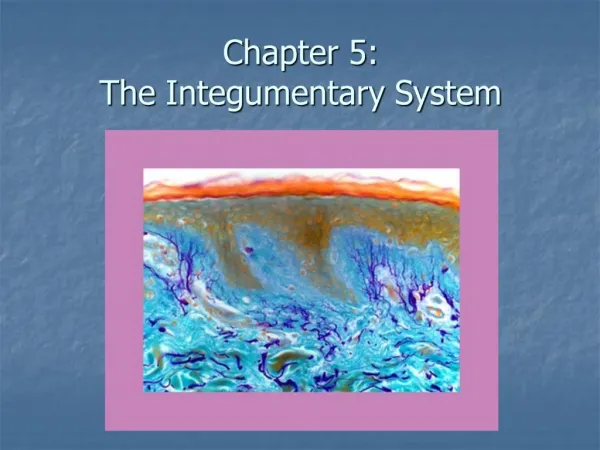
Chapter 12 Medical TerminologyandChapter 5 Body Structures: THE INTEGUMENTARY SYSTEM
FUNCTIONS OF THE INTEGUMENTARY SYSTEM – THE OUTER COVERING OF THE BODY – THE SKIN • Waterproofs body and prevents water loss • Intact skin plays important role in immune system • Receptor for the sense of touch • Screens out harmful UV rays from sun while synthesizing Vitamin D
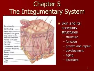
STRUCTURES OF THE INTEGUMENTARY SYSTEM • Skin (derma or cutaneous) • Epidermis • Dermis • Tissues within the dermis • Subcutaneous Layer
ASSOCIATED STRUCTURES OF THE INTEGUMENTARY SYSTEM –AND THEIR FUNCTIONS • Sebaceous glands – • secretes sebum to lubricate skin and discourage bacteria growth • Sweat glands – • Help regulate body temp and H2O content by secreting sweat – some metabolic waste secreted • Hair – • Helps control heat loss • Nails – • Protects dorsal surface of distal phalanges
THE EPIDERMIS –MADE UP OF SEVERAL LAYERS OF EPITHELIAL CELLS • Outer most layer of the skin – epidermis • Does not contain any blood vessels or connective tissue • Dependent on lower layers for nourishment • Cells are produced in lower (basal) layer and push upwards – when they reach the surface, they die and fill with keratin • Keratin: water-repellent protein • Soft keratin: primary component of epidermis • Hard keratin: found in hair and nails
THE EPIDERMIS www.aatb.org/aatbskinbank/ mission_statement.htm coolshade.tamu.edu/ skin_2.html
Cells and Layers of the Epidermis • Squamous (scalelike) epithelial tissue – • upper layer, consists of flat, scaly cells that are continuously sloughed off • Basal layer – Also contains melanocytes • Melanocytes: cells that produce and contain dark brown-black pigment (melanin) – • Type and amount of melanin determines color of skin • Melanin also protects skin against harmful UV rays of the sun
THE DERMIS – THICK LAYER OF LIVING TISSUE DIRECTLY BELOW EPIDERMIS • Contains: • Connective tissue • Blood and lymph vessels • Nerve fibers: endings receive impulses enabling body to recognize sensory stimuli like touch, temp, pain, and pressure • Hair follicles • Sebaceous and Sweat glands
TACTILE: pertaining to touch • PERCEPTION: the ability to recognize sensory stimulus www.bmb.psu.edu/.../tissues/ tissnote.htm • TISSUES WITHIN THE DERMIS – • Collagen: means glue, contains tough but flexible protein material • Also found in bone, cartilage, tendons, and ligaments • Mast Cells: respond to injury or infection by producing heparin and • histamine • Heparin: released in response to injury, is an anticoagulant • Histamine: released in response to allergens, causes itching and increased mucous secretion
THE SUBCUTANEOUS LAYER – CELLULITE = NONTECHNICAL TERM FOR SUBCUE FATTY DEPOSIT • Just below dermis • Connects skin to surface muscles • Made up of loose connective tissue and adipose (fatty) tissue • Lipocytes: fat cells, predominant in the subcutaneous layer where they manufacture and store large quantities of fat
THE SEBACEOUS GLANDS – CLOSELY ASSOCIATED WITH HAIR FOLLICLES, LOCATED IN DERMIS • Secrete sebum which is released through ducts opening into the hair follicles • Sebum lubricates skin and discourages the growth of bacteria on the skin = slightly acidic • What glands are considered part of the Integumentary system as modified sebaceous glands but are also part of the Reproductive system?? • MAMMARY GLANDS
THE SWEAT GLANDS – TINY GLANDS FOUND ON ALMOST ALL BODY SURFACES • Most numerous in palms of hands and soles of the feet, forehead, and armpits (axilla) • Pores: openings on the surface of the skin through which sweat gland ducts open • Sweat: perspiration – secreted by sweat glands • Made up of 99% water + salt + metabolic waste • Perspiration: excretion of excess H2O – cools body as sweat evaporates into air • What causes body odor associated with sweat? • INTERACTION SWEAT + BACTERIA ON SKIN
THE HAIR – FIBERS OF TIGHTLY FUSED, DEAD PROTEIN CELLS FILLED WITH HARD KERATIN • What factors determine hair color?? • Amount of melanin produced by the melanocytes that surround core of the hair shaft • Hair follicles: sacs that hold root of hair fibers • Arrector pili: (erector muscles) tiny muscle fibers attached to hair follicles that, upon contraction, cause the hair to stand up (i.e. cold or fright = goose bumps) reducing heat loss through skin
THE NAILS – UNGUIS, KERATIN PLATE COVERING DORSAL SURFACE OF DISTAL PHALANGES • Each nail consists of the following: • Nail body – translucent, made up of hard keratinized plates of epidermal cells • Nail bed – joins nail body to underlying connective tissue, nourishes the nail • Blood vessels give nail bed it’s pink color • Free edge – the portion not attached to the nail bed, extends beyond the tip of the phalanx
MEDICAL SPECIALTIES • Dermatologist • Diagnosing and treating disorders of the skin • Cosmetic surgeon – Plastic Surgeon • Restoration and repair
PATHOLOGY OF THE INTEGUMENTARY SYSTEM • Acne vulgaris: inflammatory disease with pustular eruptions of the skin in or near the sebaceous glands • Comedo: aka blackhead - sebum plug exposed to air = oxidizes • Seborrheic Dermatitis: aka dandruff – scaling of the scalp due to inflammation of upper layers of the skin
Comedo or blackheads Acne vulgaris Seborrheic dermatitis anhidrosis Sebaceous cyst
SWEAT GLAND DISORDERS • Anhidrosis: lacking sweat • Hyperhidrosis: excessive sweat • Diaphoresis: profuse, but not necessarily excessive sweating • Miliaria: heat rash/prickly heat – inflammation caused by trapped sweat
HAIR”Y “DISORDERS • Excessive Hairiness • Hirsutism: appearance of male body or facial hair patterns in the female • Abnormal hair loss • Alopecia: baldness, partial or complete • Female pattern baldness: thinning in front and on sides, sometimes on crown • Male pattern baldness: receding hairline from front to the back until only a horseshoe shaped area remains in back and temples
hirsutism folliculitis alopecia
PIGMENTATION • Albinism: deficiency or absence of pigment in skin, hair, eyes due to abnormality in production of melanin • Chloasma: mask of pregnancy – brownish colored spots on face
chloasma melanosis
SURFACE LESIONS – PATHOLOGIC CHANGE OF TISSUES DUE TO DISEASE OR INJURY • Described by appearance, location, color, and size (cm) • Contusion: does not break skin, swelling, discoloration, and pain • Ecchymosis: bruise – purple discoloration caused by hemorrhaging within the skin • Nodule:solid bump, may be felt within skin or may be raised as if it had formed below the surface and pushed upward (i.e. cyst) • Papule: solid raised skin lesion < 0.5 cm in diameter (i.e. warts, insect bites, and skin tags)
SURFACE LESIONS OF THE SKIN FLUID-FILLED LESIONS LESIONS THROUGH THE SKIN
WARNING!! • THE FOLLOWING PICTURES MAY BE DIFFICULT FOR VIEWING • VIEW AT YOUR DESCRETION
ecchymosis petechia bruise purpura contusion
dermatitis Birthmark vascular Port-wine stain Nodular skin lesions Open lesions coccidioidomycosis
Ulcers Skin ulcer post spider bite