Download
1 / 48
480 likes | 701 Views
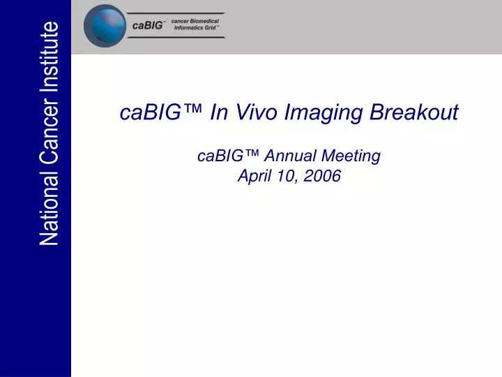
caBIG™ In Vivo Imaging Breakout caBIG™ Annual Meeting April 10, 2006. caBIG™ Annual Meeting: In Vivo Imaging Breakout Introduction by Eliot Siegel, MD In Vivo Imaging Workspace Lead. Formation of the caBIG Imaging workspace somewhat “controversial” last year

E N D
caBIG™ In Vivo Imaging Breakout caBIG™ Annual Meeting April 10, 2006
caBIG™ Annual Meeting: In Vivo Imaging BreakoutIntroduction by Eliot Siegel, MDIn Vivo Imaging Workspace Lead • Formation of the caBIG Imaging workspace somewhat “controversial” last year • Interesting mismatch between clinical use of imaging in the hospital and outpatient setting and the use of imaging in clinical trials, why is that the case? • Difficulty getting images from various sites conducting clinical trials in comparison to text based and other data • Lack of optimal quantitative tools to evaluate change in tumor over time on imaging studies • Diagnostic imaging is and will continue to play an increasingly critical role as a biomarker for disease from both a clinical and research perspective
Introduction • Unfortunately there are still major problems with images related to clinical trials • Each clinical trial group has had to reinvent the wheel which is inefficient and very expensive • ACRIN • QARC • BIRN • others • Current information systems such as PACS and the EMR are oriented toward clinical care rather than research
Introduction • Imaging Informatics is an emerging discipline in medicine and will be increasingly important in the future of cancer care • We believe that this field of informatics holds the key to making images accessible and also putting a greater degree of rigor on quantitative assessment of change over time which is critical fo the success of imaging as a biomarker • Close working relationship between NCICB (NCI-Center for Bioinformatics) NCIA (National Cancer Imaging Archive) and Imaging Workspace
caBIG™ Annual Meeting: In Vivo Imaging BreakoutIntroduction by Eliot Siegel, MDIn Vivo Imaging Workspace Lead • Goals of the workspace • Advance imaging informatics on multiple fronts • Create, optimize, and validate software tools • E.g. extensible imaging platform • Model methods to extract meaning from in vivo imaging data and establish databases to test and validate these methods
In Vivo Imaging Workspace • Structure • Eliot Siegel – Workspace Lead • Four Special Interest Groups (SIGs): • Software • Standards and Interoperability • Testbed • Vocabularies & Common Data Elements. • Fifteen Funded Subject Matter Experts (SMEs) • More than Seventy Volunteer Participants including participants from industry • Participation of NCIA and NCI/CB • Small Animal In Vivo Imaging
caBIG™ Annual Meeting: In Vivo Imaging BreakoutVocabularies and Common Data Elements Special Interest GroupLead by Curt Langlotz, MD, PhD and Daniel Rubin, MD Imaging VCDE SIG Goals • Promote, support, develop, and evaluate standards-based vocabularies, ontologies, and CDEs for radiology and allied imaging fields. • Participate in the design of the testbed and provide the vocabulary-related elements required by the testbed . • Help develop the standards for creating, storing, and retrieving image metadata and image annotations. • Harmonize VCDEs developed in the VCDE SIG with those being created by the VCDE workspace, and will develop VCDE-specific tools and resources that can be deployed on the grid to help realize the strategic vision of the caBIG effort.
Outline • Background • caBIG history • Terminology for imaging • ACRIN and imaging clinical trials • Proposed imaging VCDE projects • Structured image annotation and query software • Terminology/CDE development for imaging • Natural language processing
Clinical Trial Building Blocks Clinical Trials Tools/ Templates National Information Infrastructure Sharable Information Repositories Common Data Elements “The CII” NCI Informatics Long Range Planning, circa 1999
Importance of Common Data Collection Methods, circa 1999 • Serve as building blocks for the CII • Allow pooling of data and comparison of results among clinical trials • Facilitate enrollment of patients in clinical trials • Avoid redundant data collection (capture-once, use-many times principle) • Automate and expedite administration of clinical trials
Medical Vocabularies: Completeness for Radiology Langlotz & Caldwell, J Digit Imaging 15(1S):201, 2002
What is RadLex? 10-30 percent of these concepts are not found in SNOMED-CT • Sponsored by the Radiological Society of North America (RSNA) • 26 participating organizations • 9 committees • 92 radiologist participants • 5,308 anatomic concepts
OWL Iterative Lexicon Development Process SNOMED-CT SNOMED-CT RadLexProtégé Database RadLexweb site XML OWL RadLex base content NCI Thesaurus RadLex Lexicon Development Committees UMLS Meta-Thesaurus Lexicon Development Process
Hierarchy expandsto show results in context Term details are shown BOOP Search mirc.rsna.org/radlex/service
American College of Radiology Imaging Network(ACRIN) • NCI-funded imaging clinical trial cooperative group • Dozens of trials funded, including some very high profile trials (DMIST, NLST) • Tens of thousands of subjects • Case report forms containing thousands of potential CDEs
Proposed Imaging VCDE Projects • Structured image annotation and query (IRW) • Image meta-data standards • Image annotation and structured data capture • Image query by content from annotated image database • Data collection methods for imaging clinical trials, harmonized to RadLex and caDSR/EVS • ACRIN data collection elements • DICOM elements • The imaging “playbook”: Cancer imaging devices, procedures and protocols • Natural language processing (NLP) • Evaluation of existing tools • Adaptation or development of tools for radiology images
CAVITARY MASS Finding: mass Mass ID: 1 Margins: spiculated Length: 2.3cm Width: 1.2cm Cavitary: Y Calcified: N Spatial relationships: Abuts pleural surface; invades aorta Image Annotation and Structured Data Capture Capture data once, use it many times
Data Collection CDE Example • Please describe the margins of the mass: • Smooth • Lobulated • Irregular • Spiculated • Obscured
Vocabulary Concepts Data Collection CDE Example • Please describe the margins of the mass: • Smooth • Lobulated • Irregular • Spiculated • Obscured
Reusable Common Data Elements (CDEs) for Imaging • Create caDSR-compatible CDEs from ACRIN data collection methods • Identify CDEs specific to cancer imaging research needs • Compliant with caDSR, harmonized with RadLex and EVS • Associate atoms (terms) and molecules (CDEs) • Move from lexicon (lists) toward ontology (knowledge) • Coordinate with caBIG VCDE Workspace
The “Playbook” for Imaging in Cancer Research • Vocabulary for imaging devices, procedures, and protocols • (e.g., 7T 18-cm horizontal bore; 4.7T 33-cm bore magnet operating at 200 MHz for 1-H imaging experiments) • Common, vendor-independent language to describe experimental imaging instruments. • (e.g., “fast spin echo” vs. “turbo spin echo” MRI sequence)
Natural Language Processing • Unstructured information will always exist • Narrative radiology report archives • Peer-reviewed literature • Focused extraction from radiology report • Anatomy, findings (e.g, nodules and their descriptors), change over time • Automatic population of reporting templates • Inventory existing NLP tools • Select or develop NLP tools to fulfill requirements
Vocabulary/CDE Strategy Metadata storage formats NLP Metadata for Images Image Annotation Terminologies & CDEs Queries & Analysis Data Capture Formats & Tools Vocabularies & CDEs Data Re-Use Applications
Standards and Interoperability Special Interest GroupLead by David Channin, MD and Paul Nagy, PhD • Why Standards? • Image Standards • Workflow Standards • Annotation Standards “The great thing about standards is that there are so many to choose from.” – Dr. Andrew Tanenbaum
Why Standards? • Today, mountains of image data from clinical trials lies fallow. • The appropriate use of standards can allow re use of the image data for other purposes than the one immediate trial. • Thus enabling discovery in unanticipated ways. • Computer Assisted Diagnosis • Content Basis Image Retrieval
Image Standards • Clinical Standard Medical Images come in • DICOM (Digital Communications in Medicine) • Loads of meta data • Imaging Physics • Frame of reference • Patient/Study Information • Naming inconsistent for re use • Working with UPICT • Uniform Protocols in Clinical Trials • http://www.upict.org • RSNA, FDA, NCI, AAPM, ….. • Mapping to VCDE (Vocabulary)
Workflow Standards • How do we extract the image data from clinical environments? • Not a great deal of technical onsite expertise • Anonymization of PHI (Pseudonymization) • Electronic submission to a repository • How do we expose the data to researchers • Query of meta data • API autonomous access • Goal is to allow interoperability at multiple layers in the technology stack of the Image Platform.
Scheduled Workflow Charge Posting - Patient Info. Recon-ciliation Post-Processing Workflow Presentation of Grouped Procedures Reporting Workflow KeyImageNotes Simple Image & Numeric Reports Consistent Present-ation of Images NMImage EvidenceDocs Access to Radiology Information Portable Data for Imaging Radiology Audit Trail Option on ITI-Audit Trail and Node Authentication Basic Security Teaching File and Clinical Trial Export Workflow Comm/Query Standards IHE Radiology • LAN Based – DICOM Q/R | C-Store | GPWL • Internet based – IHE RHIO – Registry/Repository using EbXML/SOAP • Internet - DICOM WG23 utilizing OGSA
Standards and Interoperability Special Interest GroupLead by David Channin, MD and Paul Nagy, PhD Annotations and Image Markup – In conjunction with Vocabulary Courtesy Dr. David Clunie
Annotations and Markup • Goal is to create a knowledge representation (OWL) for annotations in markup to enable semantic web applications. • Provide practical presentation states in DICOM Structured Reports and XML RIDER. • Create tools in the Imaging Platform to author this markup.
Imaging Software SIG: Goals and Objectives • The goal of the Software SIG is to create and adapt open source software tools to promote and enhance the use of imaging in cancer research. The SIG will focus on tools for image acquisition, management and analysis for use in clinical trials. • specifically tools for enhancing lesion detection, characterization and change determination. • The SIG will define requirements for these projects and write requirements specifications and/or white papers. • The SIG will define use cases and test plans for each project and guide and track the development team that is tasked with implementation. • The SIG will participate in the validation of software and/or algorithms resulting from each project using the IVI test bed
Viewing, Annotation and Analysis Software • To facilitate the increased use of imaging based end points in clinical trials the SIG has identified the need for an easily extensible open source platform to support image analysis and visualization. • To address this need a development program will been undertaken to create an eXtensible Imaging Platform (XIP)
eXtensible Imaging Platform • The XIP is a • Collection of software classes, algorithms and sample applications for building imaging applications valuable to research • Method for rapidly prototyping "medical imaging workstation" applications from a re-usable, extensible set of modular elements • Researchers will be able to rapidly develop and evaluate new approaches to medical imaging problems, and use them in a translational research setting. • Grid technology in general, and caGrid in particular, makes it possible to let users to choose between grid components and locally available components. • Analytic services (CAD algorithms, algorithms for quantifying changes in consecutive imaging studies, algorithms associated with a 3-D visualization pipeline etc). • Data sources might or might not be DICOM based. • Both data and algorithms can be physically distributed.
Current Status • Imaging Software SIG has developed a requirements specification for the XIP • An RFP has been drafted • The SIG is working to define appropriate milestones and demonstration projects
Change Detection & Analysis • The In Vivo Imaging Workspace is assessing current status of change detection & analysis technology for cancer imaging • Detecting and quantifying change in lesions over time represents a critical unmet need in the Cancer Research Community
Baseline Follow-up
Baseline Follow-up
Change Analysis and Validation • Working on definition of SIG’s role in larger NCI activity. It has been suggested by NCI that a major contribution would be the development of basic change analysis algorithms, and evaluation methods. • Algorithms for “binary outcome” determination and for change quantification • Databases with known truth for validation studies • Databases containing multiple segmentation results on the same images using different approaches • Utilize the “plug-in” application interface for the XIP to provide a “sand box” in which algorithms may be implemented and evaluated
Testbed Special Interest GroupLead by Joel Saltz, MD, PhD and Stephan Erberich PhD SIG Goals • Design and implement core middleware compliant with caGrid, DICOM and IHE • Addresses the need for high performance data transport on the grid, and dynamic algorithm deployment to reduce the need to data movement. • Develop software development environments to help developers use middleware to develop applications • Work with cooperative groups to leverage testbed capabilities in support of translational research • Responsibility for coordination of GridCad application
Testbed Special Interest GroupLead by Joel Saltz, MD, PhD and Stephan Erberich PhD Testbed: Consists of vivo imaging caGrid standards, reference middleware stack implementation supporting grid based applications. The testbed is designed to support each individual application as well as to demonstrate interoperability between applications Testbed Projects: Middleware, Coordination of Application Projects, Cooperative group outreach
Middleware Testbed for multi-center clinical trials:Cooperative Cancer Group use case scenario • ACQUISITION • Image acquisition and handling at trial site (Image transfer techniques, HIPAA, firewalls, MIRC) • Quality assurance • REVIEW AND ANALYSIS • Image Warehousing, access control, and central review • Access to correlative and image meta data via caGrid • Annotation and Markup in caGrid • Quantitative analytic tools • DISCOVERY • Integrated caGrid supported discovery of image, molecular, pathology information
Testbed development focus application: gridCAD — A Novel Use of Grid Computing to Support Human Markup and Execution of Multiple CAD Systems Tony Pan, Joel Saltz, Tahsin Kurc, Stephen Langella, Shannon Hastings, Scott Oster, Ashish Sharma, Metin Gurcan Department of Biomedical Informatics The Ohio State University Medical Center, Columbus OH Eliot Siegel, Khan M. Siddiqui University of Maryland School of Medicine, Baltimore, MD
Benefits • Facilitate research and clinical decision support with large number of subjects and multiple CAD algorithms. • Parameter studies, clinical and preclinical trials, etc • Provide a client to support remote human markup of nodules • Enable better algorithm development and validation through the use of many distributed, shared image datasets • Support remote algorithm execution – reduce data transfer and avoid the need to transmit PHI • Reduce overall processing time and algorithm development cycle through remote compute resource recruitment and CAD compute farms • Scalable and open source — caGrid 1.0
gridCAD Architecture Expose algorithms, human markup and image data as caGrid Services
Future Direction • Location independence • Move algorithms to data • Move data to algorithms • Move both data and algorithms to compute servers • Currently supported – ongoing collaborations to deploy these capabilities • Security and Privacy • Encryption and Just-In-Time anonymization for the image data services • Scaling and Deployment • High performance image transfer mechanisms • Greater number and variety of CAD vendors • Additional application areas, including CAD for other diseases and in vitro image analysis
COG/NANT Cooperative Group Application • COG Phase-I Consortium (23 medical center) and NANT (14 medical centers) are now actively engaged in the caBIG testbed. • Grid based analysis of perfusion imaging studies: DCE-MRI analysis deployed as an analytic service • Grid based evaluation of joint prognostic value of perfusion studies, pathology, molecular clinical data
In Vivo Imaging Workspace involvement in RSNA 2006Eliot Siegel, MD, 6 Workstations: SIG 1 Testbed Architecture 3 Workstations: SIG 3 Vocabulary 2 Workstations: caBIG demo 1 spy, Rembrandt 2 Workstations: NCI CIP demo Projects IDRi Theme Park Directory & Purpose 6 Workstations: SIG 2 Software 3 Workstations: SIG 4 Standards 2 Workstations: NCIA RIDER 4 Workstations: Allies Pharma Device ?CRO
Q&A and Wrap UpLead by Eliot Siegel, MD, In Vivo Imaging Workspace Lead • Engage in projects that further the strategic goals of the Imaging Workspace and caBIG™ program. • Identify synergies with the other caBIG™ workspaces. • Partner with external organizations within the caBIG™ community, (ex.; ACRIN, NCIA), to further Imaging Workspace and caBIG™ program goals