Download
1 / 27
270 likes | 278 Views
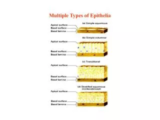
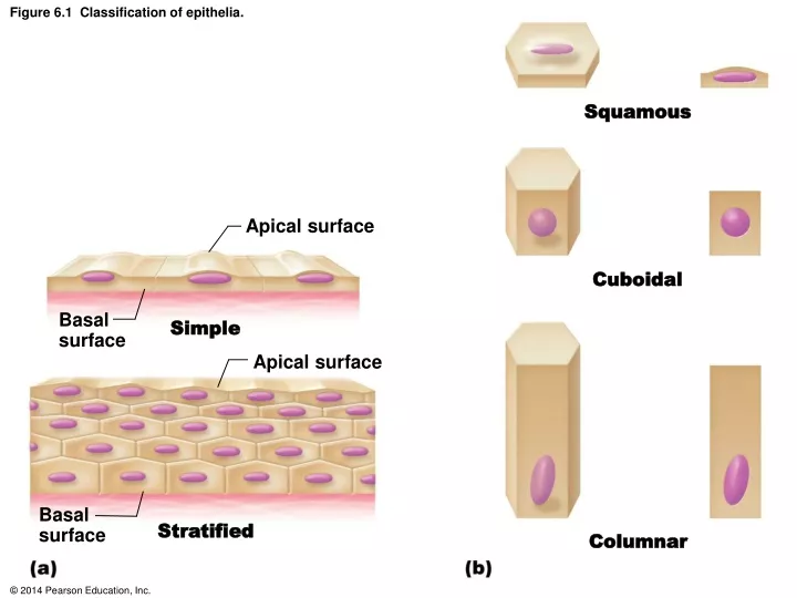
Squamous. Figure 6.1 Classification of epithelia. Apical surface. Cuboidal. Basal surface. Simple. Apical surface. Basal surface. Stratified. Columnar. Figure 6.2 Formation of endocrine and exocrine glands from epithelial sheets. Lumen of gland. Secretory cells. Exocrine gland.

E N D
Squamous Figure 6.1 Classification of epithelia. Apical surface Cuboidal Basalsurface Simple Apical surface Basalsurface Stratified Columnar
Figure 6.2 Formation of endocrine and exocrine glands from epithelial sheets. Lumen of gland Secretory cells Exocrine gland Epithelium Area ofatrophied duct Capillaries Cord ofinvaginatingepithelial cells Secretory cells Endocrine gland
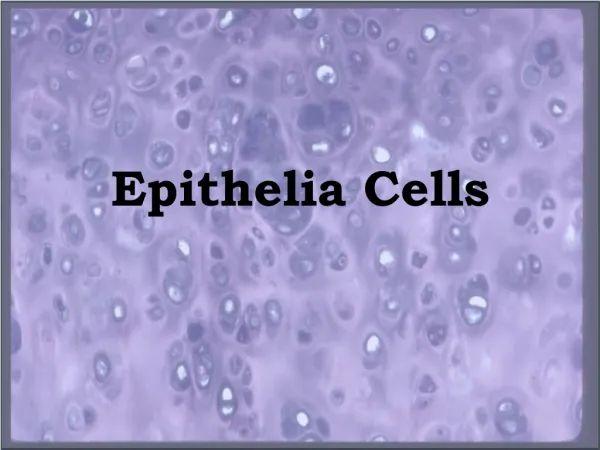
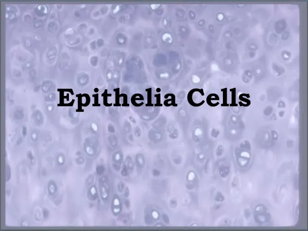
Figure 6.3a Epithelial tissues. Simple squamous epithelium Description: Single layer of flattened cellswith disc-shaped central nuclei and sparsecytoplasm; the simplest of the epithelia. Air sacs oflung tissue Nucleiof squamousepithelialcells Function: Allows materials to pass bydiffusion and filtration in sites whereprotection is not important; secreteslubricating substances in serosae. Location: Kidney glomeruli; air sacs oflungs; lining of heart, blood vessels, andlymphatic vessels; lining of ventral bodycavity (serosae). Photomicrograph: Simple squamous epitheliumforming part of the alveolar (air sac) walls (140).
Figure 6.3b Epithelial tissues. Simple cuboidal epithelium Description: Single layer of cubelikecells with large, spherical centralnuclei. Simplecuboidalepithelialcells Function: Secretion and absorption. Basement membrane Location: Kidney tubules; ducts andsecretory portions of small glands;ovary surface. Connectivetissue Photomicrograph: Simple cuboidal epitheliumin kidney tubules (430).
Figure 6.3c Epithelial tissues. Simple columnar epithelium Description: Single layer of tall cells withround to oval nuclei; some cells bear cilia;layer may contain mucus-secretingunicellular glands (goblet cells). Goblet cells Mucussecretion Function: Absorption; secretion of mucus,enzymes, and other substances; ciliatedtype propels mucus (or reproductive cells)by ciliary action. Location: Nonciliated type lines most ofthe digestive tract (stomach to rectum),gallbladder, and excretory ducts of someglands; ciliated variety lines small bronchi,uterine tubes, and some regions of theuterus. Microvilli(brush border) Photomicrograph: Simple columnar epithelium containinggoblet cells from the small intestine (640).
Figure 6.3d Epithelial tissues. Pseudostratified columnar epithelium Description: Single layer of cells of differingheights, some not reaching the free surface;nuclei seen at different levels; may containmucus-secreting goblet cells and bear cilia. Mucus ofgoblet cell Cilia Function: Secretes substances, particularlymucus; propulsion of mucus by ciliary action. Pseudo-stratifiedepitheliallayer Location: Nonciliated type in male’ssperm-carrying ducts and ducts of largeglands; ciliated variety lines the trachea,most of the upper respiratory tract. Basementmembrane Trachea Photomicrograph: Pseudostratified ciliatedcolumnar epithelium lining the human trachea (530).
Figure 6.3e Epithelial tissues. Stratified squamous epithelium Description: Thick membrane composed ofseveral cell layers; basal cells are cuboidal orcolumnar and metabolically active; surfacecells are flattened (squamous); in thekeratinized type, the surface cells are full ofkeratin and dead; basal cells are active inmitosis and produce the cells of the moresuperficial layers. Stratified squamous epithelium Function: Protects underlying tissues inareas subjected to abrasion. Nuclei Location: Nonkeratinized type forms themoist linings of the esophagus, mouth, andvagina; keratinized variety forms theepidermis of the skin, a dry membrane. Basementmembrane Connective tissue Photomicrograph: Stratified squamous epitheliumlining the esophagus (280).
Figure 6.3f Epithelial tissues. Stratified cuboidal epithelium Description: Generally twolayers of cubelike cells. Basement membrane Cuboidalepithelialcells Function: Protection Location: Largest ducts ofsweat glands, mammaryglands, and salivary glands. Duct lumen Photomicrograph: Stratified cuboidalepithelium forming a salivary glandduct (290).
Figure 6.3g Epithelial tissues. Stratified columnar epithelium Description: Several cell layers;basal cells usually cuboidal;superficial cells elongatedand columnar. Basementmembrane Stratifiedcolumnarepithelium Function: Protection; secretion. Location: Rare in the body; smallamounts in male urethra and inlarge ducts of some glands. Connectivetissue Urethra Photomicrograph: Stratified columnar epitheliumlining of the male urethra (290).
Figure 6.3h Epithelial tissues. Transitional epithelium Description: Resembles both stratifiedsquamous and stratified cuboidal; basalcells cuboidal or columnar; surface cellsdome shaped or squamouslike, dependingon degree of organ stretch. Transitionalepithelium Function: Stretches readily and permitsdistension of urinary organ by containedurine. Location: Lines the ureters, urinarybladder, and part of the urethra. Basementmembrane Connectivetissue Photomicrograph: Transitional epithelium lining the urinarybladder, relaxed state (365); note the bulbous, or rounded,appearance of the cells at the surface; these cells flatten andbecome elongated when the bladder is filled with urine.
Figure 6.4 Areolar connective tissue: A prototype (model) connective tissue. Cell types Extracellularmatrix Ground substance Macrophage Fibers • Collagen fiber • Elastic fiber • Reticular fiber Fibroblast Lymphocyte Fat cell Capillary Mast cell Neutrophil
Figure 6.5a Connective tissues. Embryonic connective tissue: Mesenchyme Description: Embryonic connectivetissue; gel-like ground substancecontaining fibers; star-shapedmesenchymal cells. Fibers Function: Gives rise to all otherconnective tissue types. Groundsubstance Location: Primarily in embryo. Mesenchymalcell Photomicrograph: Mesenchymal tissue, an embryonicconnective tissue (627); the clear-appearing background isthe fluid ground substance of the matrix; notice the fine,sparse fibers.)
Figure 6.5b Connective tissues. Connective tissue proper: loose connective tissue, areolar Description: Gel-like matrix with allthree fiber types; cells: fibroblasts,macrophages, mast cells, and somewhite blood cells. Collagenfibers Function: Wraps and cushions organs;its macrophages phagocytize bacteria;plays important role in inflammation;holds and conveys tissue fluid. Fibroblastnuclei Location: Widely distributed underepithelia of body, e.g., forms laminapropria of mucous membranes;packages organs; surrounds capillaries. Elasticfibers Epithelium Photomicrograph: Areolar connective tissue, a soft packagingtissue of the body (365). Laminapropria
Figure 6.5c Connective tissues. Connective tissue proper: loose connective tissue, adipose Description: Matrix as in areolar,but very sparse; closely packedadipocytes, or fat cells, havenucleus pushed to the side bylarge fat droplet. Function: Provides reserve fuel;insulates against heat loss;supports and protects organs. Vacuolecontainingfat droplet Location: Under skin; aroundkidneys and eyeballs; withinabdomen; in breasts. Nuclei offat cells Photomicrograph: Adipose tissue from thesubcutaneous layer under the skin (110).
Figure 6.5d Connective tissues. Connective tissue proper: loose connective tissue, reticular Description: Network of reticularfibers in a typical loose groundsubstance; reticular cells lie onthe network. Function: Fibers form a softinternal skeleton (stroma) thatsupports other cell types,including white blood cells, mastcells, and macrophages. White blood cell(lymphocyte) Location: Lymphoid organs(lymph nodes, bone marrow,and spleen). Reticularfibers Spleen Photomicrograph: Dark-staining network ofreticular connective tissue fibers forming theinternal skeleton of the spleen (350).
Figure 6.5e Connective tissues. Connective tissue proper: dense connective tissue, dense regular Description: Primarily parallelcollagen fibers; a few elastic fibers;major cell type is the fibroblast. Function: Attaches muscles to bonesor to muscles; attaches bones to bones;withstands great tensile stress whenpulling force is applied in one direction. Collagenfibers Location: Tendons, mostligaments, aponeuroses. Nuclei offibroblasts Shoulderjoint Ligament Photomicrograph: Dense regular connective tissuefrom a tendon (590). Tendon
Figure 6.5f Connective tissues. Connective tissue proper: dense connective tissue, elastic Description: Dense regular connectivetissue containing a high proportion ofelastic fibers. Function: Allows recoil of tissuefollowing stretching; maintainspulsatile flow of blood througharteries; aids passive recoil of lungsfollowing inspiration. Elasticfibers Location: Walls of large arteries;within certain ligaments associatedwith the vertebral column; within thewalls of the bronchial tubes. Aorta Heart Photomicrograph: Elastic connective tissue in thewall of the aorta (250).
Figure 6.5g Connective tissues. Connective tissue proper: dense connective tissue, dense irregular Description: Primarily irregularlyarranged collagen fibers; some elasticfibers; major cell type is the fibroblast. Nuclei offibroblasts Function: Able to withstand tensionexerted in many directions; providesstructural strength. Location: Fibrous capsules of organsand of joints; dermis of the skin;submucosa of digestive tract. Collagenfibers Fibrousjointcapsule Photomicrograph: Dense irregular connectivetissue from the dermis of the skin (210).
Figure 6.5h Connective tissues. Cartilage: hyaline Description: Amorphous but firm matrix;collagen fibers form an imperceptiblenetwork; chondroblasts produce thematrix and when mature (chondrocytes)lie in lacunae. Chondrocytein lacuna Function: Supports and reinforces;serves as resilient cushion; resistscompressive stress. Location: Forms most of the embryonicskeleton; covers the ends of long bonesin joint cavities; forms costal cartilagesof the ribs; cartilages of the nose,trachea, and larynx. Matrix Costalcartilages Photomicrograph: Hyaline cartilage from a costalcartilage of a rib (470).
Figure 6.5i Connective tissues. Cartilage: elastic Description: Similar to hyalinecartilage, but more elasticfibers in matrix. Function: Maintains the shapeof a structure while allowinggreat flexibility. Chondrocytein lacuna Location: Supports the externalear (auricle); epiglottis. Matrix Photomicrograph: Elastic cartilage from thehuman ear auricle; forms the flexible skeletonof the ear (510).
Figure 6.5j Connective tissues. Cartilage: fibrocartilage Description: Matrix similar tobut less firm than that in hyalinecartilage; thick collagen fiberspredominate. Function: Tensile strength withthe ability to absorb compressiveshock. Location: Intervertebral discs;pubic symphysis; discs of kneejoint. Chondrocytesin lacunae Intervertebraldiscs Collagenfiber Photomicrograph: Fibrocartilage of an intervertebraldisc (160). Special staining produced the blue color seen.
Figure 6.5k Connective tissues. Bones (osseous tissue) Description: Hard, calcified matrixcontaining many collagen fibers;osteocytes lie in lacunae. Verywell vascularized. Centralcanal Function: Bone supports andprotects (by enclosing); provideslevers for the muscles to act on;stores calcium and other mineralsand fat; marrow inside bones is thesite for blood cell formation(hematopoiesis). Lacunae Lamella Location: Bones Photomicrograph: Cross-sectional view of bone (175).
Figure 6.5l Connective tissues. Blood Description: Red and whiteblood cells in a fluid matrix(plasma). Plasma Neutrophil Function: Transport ofrespiratory gases, nutrients,wastes, and other substances. Red bloodcells Location: Contained withinblood vessels. Lymphocyte Photomicrograph: Smear of human blood (1000);two white blood cells (neutrophil and lymphocyte) areseen surrounded by red blood cells.
Figure 6.6 Nervous tissue. Nervous tissue Description: Neurons are branchingcells; cell processes that may be quitelong extend from the nucleus-containingcell body; also contributing to nervoustissue are nonexcitable supporting cells. Nuclei ofsupportingcells Cell body Neuron processes Axon Dendrites Cell bodyof a neuron Function: Neurons transmit electricalsignals from sensory receptors and toeffectors (muscles and glands); supportingcells support and protect neurons. Neuronprocesses Location: Brain, spinalcord, and nerves. Photomicrograph: Neurons (370)
Figure 6.7a Muscle tissues. Skeletal muscle Description: Long, cylindrical,multinucleate cells; obviousstriations. Part ofmusclefiber (cell) Function: Voluntary movement;locomotion; manipulation of theenvironment; facial expression;voluntary control. Nuclei Location: In skeletal musclesattached to bones or occasionallyto skin. Striations Photomicrograph: Skeletal muscle (approx. 550).Notice the obvious banding pattern and thefact that these large cells are multinucleate.
Figure 6.7b Muscle tissues. Cardiac muscle Description: Branching, striated,generally uninucleate cells thatinterdigitate at specialized junctionscalled intercalated discs. Nucleus Intercalateddiscs Function: As it contracts, itpropels blood into the circulation;involuntary control. Striations Location: The walls of the heart. Photomicrograph: Cardiac muscle (775);notice the striations, branching of cells, andthe intercalated discs.
Figure 6.7c Muscle tissues. Smooth muscle Description: Spindle-shaped cellswith central nuclei; no striations;cells arranged closely to formsheets. Smooth muscle cell Function: Propels substances orobjects (foodstuffs, urine, a baby)along internal passageways;involuntary control. Nucleus Location: Mostly in the walls ofhollow organs. Photomicrograph: Smooth muscle cells (265).