Download
1 / 66
660 likes | 949 Views
Digital Radiography. Or Things they never tell you when the equipment is installed REV 11/2008. Computed Radiography. Fundamentals of Computerized Radiography. Imaging Plates. Latitude and Contrast Independent functions

E N D
Digital Radiography Or Things they never tell you when the equipment is installed REV 11/2008
Computed Radiography Fundamentals of Computerized Radiography
Imaging Plates • Latitude and Contrast • Independent functions • Contrast is dependent on look-up table (LUT) and final image processing • Image appearance not as closely related to exposure factor selection as F/S was • Except when extremely out of range
EDR • Exposure Data Recognition • When laser scans it is looking for area of plate that has exposure • Some read from center out and look for two sides of collimation • Works best when image centered
Histogram • Identifies all densities on plate • A graph that displays signal value • x-axis related to amount of exposure • y-axis displays number of pixels for each exposure • Series of peaks and valleys • Pattern varies for each body part
Histogram Location • Example of a chest histogram • X-ray energy and absorption effects • Low kVp, large differences (high contrast) • High kVp, small differences (low contrast) • Different energies superimposed • Different doses (mAs), same kVp
Darker Lighter Histogram showing pixel values in an image. The pixel values in gray are on the horizontal with the total number for each on the vertical.
Histogram Analysis • Collimation is very important • If plate reader cannot find collimated edges then all the exposure on plate will be included in the histogram • Histogram from plate is compared to body part histogram stored in computer
LUT • Look Up Table (LUT) • Each anatomic area has a LUT • Used to adjust contrast and density • Other terms that may be used for this • Contrast rescaling • Contrast processing • Gradation processing • Tone scaling
LUT • The image data from the histogram is rescaled for application of the LUT • The LUT maps the adjusted data through a “S” curve that is similar to an H & D curve • The result is an image that has the correct contrast and brightness (density)
LOOK UP TABLE (LUT) Black Saturation White Saturation Linear LUT Black Shirt Facial Tones * No Detail in Black Areas * High Contrast * Only Detail in White Areas can be seen * No Detail in White Areas * Low Contrast * Only Detail in Black Areas can be seen
Exposure Indicators • Imaging plates get a signal from the exposure they receive • The value of the signal is calculated from the region identified as the anatomy of interest • The signal for the plate is an average of all signals given to the plate
The total signal is not a measure of the dose to the patient but indicates how much radiation was absorbed by the plate • A 1 mR exposure will give • Fuji S# 200 • Kodak EI 2,000 • Agfa 200 speed lgm reference value for site
EXPOSURE VALUES • Exposure indicator • Plates sensitive to 0.1 mR – 100 mR • “S” number for Fuji • S number inverse to exposure • S=2 (100 mR), S=200 (1 mR) • Kodak uses exposure index • 2000 (1 mR), 3000 (10 mR)
Exposure Values • Agfa has ADC dose monitoring • Stored reference dose for each exam • Based on range • Compares image obtained to reference • Indicates significant deviation • Notes lgM, not patient dose, plate dose • Doubling dose increases lgM by 0.3 (log)
Exposure Values • Agfa • lgM, log gradient mean • Double dose is 0.3 since it is log • Have stored reference dose for exams • Set for hospital, each image compared to it • Speed class, 200 or 400 • Tells relative amount of light to be read from plate • Allows electronics to be optimized to this range for better image data set
Agfa • Agfa uses a speed system • Here is a hand taken at 50, 200 and 400 speeds • The mAs was changed for each one • Lgm is the same for all
Using Exposure Numbers • Fuji, if appropriate # is 200 then • At 400, too light, double mAs for 200 • At 100, too dark, half mAs for 200 • Agfa, if appropriate # is Lgm 2.2 • At 1.9, too light, double mAs for 2.2 • At 2.5, to dark, half mAs for 2.2 • Kodak, if appropriate # is 1800 • At 1500, too light, double mAs for 1800 • At 2100, to dark, half mAs for 1800
S# 47 S# 86 S# 16
S# 8,357 S# 12,361 lat CXR
Exposure Numbers • The exposure numbers can only be used if all other parameters are correct • Centering to plate • Collimation • Position over AEC, look at mAs readout to determine if poor positioning caused light or dark image
Same technique, different centering and collimation S# 592 S# 664
Exposure Values • Each system has range of values for appropriate exposure for part • The range used by vendor is very broad • Each facility should develop its own exposure range taking into account • Radiologist preference • ALARA
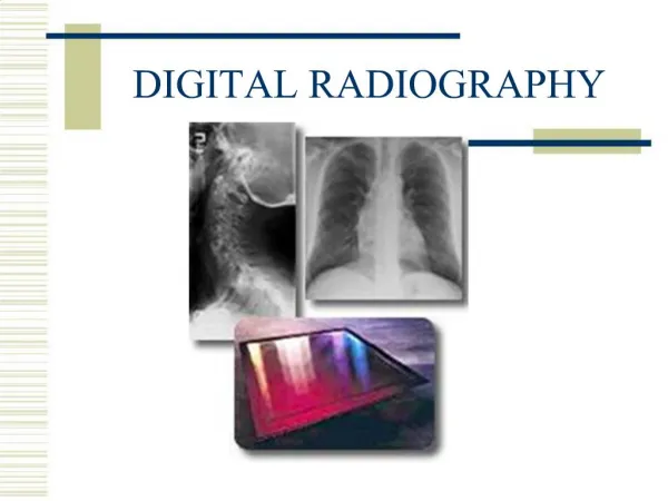
IMAGE CAPTURE CR • PSP – photostimulable phosphor plate • REPLACES FILM IN THE CASSETTE DR – NO CASSETTE – PHOTONS • CAPTURED DIRECTLY • ONTO A TRANSISTOR • SENT DIRECTLY TO A MONITOR
DR • TFT, thin film transistor • Used under silicon or selenium • Collects charge then sends out as signal to computer • Releases e- line by line
CR – PSP plate • Stimulated by a RED LIGHT • Energy is RELEASED in a form of BLUE light • LIGHT captured by PMT – • changed to a digiial signal
How CR works • Released light is captured by a PMT (photo multiplier tube) • This light is sent as a digital signal to the computer • The intensity (brightness) of the light – correlates to the density on the image
Densities of the IMAGE • The light is proportional to amount of light received • digital values are then equivalent (not exactly the same) to a value of optical density (OD) from a film, at that location of the image
ADVANTAGE OF CR/DR • CHANGES MADE TO IMAGE • AFTER THE EXPOSURE • CAN ELIMINATE THE NEED TO REPEAT THE EXPOSURE
ADVANTAGE OF CR/DR vs FS • Rapid storage • retrieval of images NO LOST FILMS! • PAC (storage management) • Teleradiology - long distance transmission of image information • Economic advantage - at least in the long run?
CR/DR VS FILM/SCREEN • FILM these can not be modified once processed • If copied – lose quality • DR/CR – print from file – no loss of quality
“no fault” TECHNIQUES F/S: RT must choose technical factors (mAs & kvp) to optimally visualize anatomic detail CR: the selection of processing algorithms and anatomical regions controls how the acquired latent image is presented for display • HOW THE IMAGE LOOKS CAN BE ALTERED BY THE COMPUTER – EVEN WHEN “BAD” TECHNIQUES ARE SET
DR • Initial expense high • very low dose to pt – • image quality of 100s using a 400s technique • Therfore ¼ the dose needed to make the image
Histogram Analysis • A histogram is a plot of gray scale value • vs. the frequency of occurrence • (# pixels) of the gray value in the image
HISTOGRAM– a bar graph depicting the density distribution (in numerical values) of the imaging plate • ALGORITHM – a set of mathematical values used to solve a problem or find an average