Download
1 / 32
320 likes | 325 Views
This article discusses the role of the endoplasmic reticulum (ER) in protein disulfide formation and redox signaling. It explores the various pathways involved in disulfide bond formation and the generation of reactive oxygen species (ROS) within the ER. The article also highlights the relevance of ER stress and unfolded protein response in various diseases and pathogenic mechanisms.

E N D
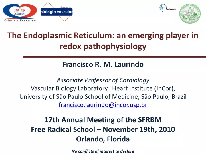
The Endoplasmic Reticulum: an emerging player in redox pathophysiology Francisco R. M. Laurindo Associate Professor of Cardiology Vascular Biology Laboratory, Heart Institute (InCor), University of São Paulo School of Medicine, São Paulo, Brazil francisco.laurindo@incor.usp.br 17th Annual Meeting of the SFRBM Free Radical School – November 19th, 2010 Orlando, Florida No conflicts of interest to declare
Role of disulfide bonds in proteins • Foldingstabilizationandfunctionalcapacitationofmembrane, cellsurfaceorsecretedproteins Also....: • Controlofpeptideloading in the MHC • Thiol-mediated ER retentionof some proteins (ex: Ero1) • Thrombogenicity The endoplasmic reticulum is the major site for protein disulfide introduction and isomerization in eukaryotes
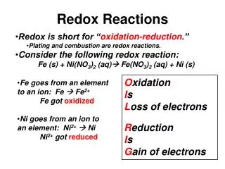
Ancient disulfide relay systems have terminal electron sinks for reduced molecular oxygen... • Bacterial periplasm: • Main components: DsbA/DsbB • Electron sink: ubiquinone / terminal membrane oxidase • Mitochondrial intermembrane space: • Main components: Mia40 / Erv1 (FAD) • Electron sink: cytochrome c / cytochrome oxidase ... and the final product is H2O
FAD-binding pocket of Ero 1 (Chu Y et al, BBRC 2009) Ero1p oxidizes PDI and transfer electrons to oxygen, generating hydrogen peroxide: catalytic and shuttle disulfides FAD hidrophobic residues (Tavender TJ and Bulleid NJ. Antiox Redox Signal 2010; 13: 1177-87)
Characteristics of PDI (-SH pKa 6.2; ’o-180 mV) • PDI is a thioredoxin superfamily dithiol-disulfide oxidoreductase chaperone from the endoplasmic reticulum. The PDI family has >17 known proteins. • PDI may complex with other chaperones: Grp78, Grp94, P5, ERdj3, cyclophilin B, Erp72, Grp170, UGGT, SDF2 (Meunier et al 2002) • PDI may act as thiol reductase or oxidase, but the hallmark of PDI family is the isomerase activity: PDI PDI PDI Laurindo et al 2008 Modif. from Hatahet and Ruddock 2009 PDI PDI • PDI also has chaperone activity, which is in itself independent on thiol groups, but is synergic with disulfide transfer.
Regulatory disulfides in Ero1p * *analogous disulfides exist for mammalian Ero1α Tavender TJ and Bulleid NJ. Antiox Redox Signal 2010; 13: 1177-87
Genetic deletion of Ero1β promotes glucose intolerance, but mice with double Ero1 β/Ero1α deletion are viable and have no additional phenotype :another challenge to the concept of Ero1-PDI as the only disulfide-generating system Zito et al. J Cell Biol 2009; 188: 821-32)
Summary of pathways for protein disulfide formation Disulfide bond formation in proteins ascorbate/ DHA vit K vit K-OR Sox family (QSOX) PDI family GSSG Ero1 family O2 H2O2 via regulatory disulfides outside the ER within the ER
Endoplasmic reticulum: an underappreciated source of ROS • Quantitation • Superficial calculation suggests up to 25% of cell ROS (Tu and Weissman . J Cell Biol 2004) • Low antioxidant concentration • Very low or nonexistent SODs, catalase or glutathione reductase; little reference to GPx(s), Trx/TrxR; glutathione transferase only in hepatocytes (for references, see Santos CX et al, ARS 2009) • Secreted ER-specific PrxIV forms decamers with no obvious antioxidant activities(Tavender et al 2008), although some reports do suggest antioxidant effect(Okado-Matsumoto et al, 2000) • ER: the major source of GSSG in the cell (in yeast, via Ero1p – Cuozzo and Kaiser, Nature Cell Biol 1999)
The ER lumen is a relevant source of H2O2:studies with HyPer sensor targeted to cytosol, mitochondria, nucleus, plasma membrane, ER lumen or ER cytosolic surface Enyedi B et al. Antiox Redox Signal 2010; 13: 721
Ero1α regulates ER levels of H2O2 Hyper-ER fluorescence Enyedi B et al. Antiox Redox Signal 2010; 13: 721
Un/misfolded protein accumulation at the ER lumen Endoplasmic reticulum (ER) stress Unfolded protein response (UPR) 1- Arrest in mRNA translation and synthesis of new protein 2- ER expansion 3- Overexpression of ER chaperones 4- Apoptosis The 4 “A”s of the UPR (Rutkowski & Kaufman, TBS 2007): Activation Acute response Adaptation Apoptosis
Many diseases/pathogenetic mechanisms are associated with ER stress signaling • Tumors(Herr et al, Blood 2001) • Parkinson´s disease and Neurodegenerative Diseases (Paschen & Doutheil, JCBFM 1999; Zhao & Ackerman, Curr Opin Cell Biol 2006) • -1 antitrypsin deficiency (Lawless et al, J Immunol 2004) • Viral infection (He, Cell Death Differ 2006) • Inflammation (for a review, see Zhang K and Kaufman R, Nature 2008) • Atrial fibrillation (Vitadello et al, Circulation 2001) • Cardiac hypertrophy and failure (Okada et al, Circulation 2004) • Hyperhomocysteinemia(Werstuck et al, J Clin Invest 2001) • Obesity, diabetes and insulin resistance(Ozcan et al, Science 2004 & 2006; for a review, see Scheuner and Kaufman , Endocr Rev 2008) • Atherosclerosis (Zhou et al, Circulation 2005) • Cholesterol toxicity / lipid metabolism (reviewed by Marciniak and Ron, Physiol Rev 2006)
UPR signaling induced by ER stress ( modif. from Santos et al, ARS 2009) Proapoptotic signaling Proadaptative signaling ER-associated mRNA degradation ER lumen kinase endonuclease IRE1 IRE1 JNK/c-jun XBP1 mRNA splicing P P Caspase-12 Caspase-9 Caspase-3 cleavage @ Golgi ? ATF6 ATF6 PERK Nrf2 P antioxidant genes ? ↑CHAPERONE eIF2α eIF2α P grp78 grp94 calreticulin PP1c arrest in protein translation GADD34 ATF4 CHOP/GADD153 Genes for:aminoacid sufficiency, antioxidants, metabolic adaptations, ERAD Apoptosis
Main operational markers of the UPR • ER stress sensors • IRE1 phosphorylation • PERK phosphorylation • ATF6 cleavage, nuclear migration • UPR pathways • eIF2α phosphorylation • XBP1 mRNA (and protein) splicing • ATF4 nuclear expression • CHOP/GADD153 nuclear expression • Expression of KDEL-bearing chaperones: Grp78 (BIP), Grp94, calreticulin, Orp150
The UPR generates ROS at several levels and ROS feedback on the UPR (for a review of many other examples, see Santos CX et al, ARS 2009) 100 Tunicamycin 2µg/ml; 16h 80 Basal 60 Superoxide generation – EOH/DHE Δ% Tn vs. Basal Grp94 40 Grp78 20 -actin - + - + Catalase cDNA siRNA caspase12 Control siRNA Scr GADD34 GADD34ΔC
Futile Ero1/PDI/glutathione cycles of attempted folding lead to ER-dependent ROS generation and GSH consumption during ER stress O2 H2O2 FADH2 FAD+ Ero-1 Ero-1 SH SH S S PDI PDI SH SH S S Protein Protein SH SH S S GSH GSSG
Futile Ero1/PDI/glutathione cycles of attempted folding lead to ER-dependent ROS generation and GSH consumption during ER stress O2 H2O2 FADH2 FAD+ Ero-1 Ero-1 SH SH S S PDI PDI SH SH S S Misfolded protein substrates SH SH S S Non-native disulfides GSH GSSG
Futile Ero1/PDI/glutathione cycles of attempted folding lead to ER-dependent ROS generation during ER stress: caveats • Constitutively increased Ero1 overexpression does not lead to detectable oxidant generation, at least in unstressed cells (Sevier et al. Cell 2007). • In vivo 1:1 stoichiometry of H2O2 production via Ero1 is unclear. • Rate constants of Ero1/PDI/protein/glutathione thiol exchanges may be quite low. Example: 182 M-1.s-1 for DsbA vs. GSH (Hu et al, J Prot Chem 1999). • Electron acceptors other than oxygen can potentially support Ero1p oxidation in some instances (Tu et al, Science 2000). • Free FAD levels support Ero1/PDI/protein oxidation (Papp et al, BBRC 2005) and strongly affect Ero1p activity (although less so for Ero1α – Wang et al JBC 2009). Little is known regarding FAD sufficiency in the ER.
Futile Ero1/PDI/glutathione cycles of attempted folding lead to ER-dependent ROS generation during ER stress: caveats • ER seems underoxidized during yeast UPR. Some ER underoxidation still detected during ER stress due to transfection of cys-free misfolded substrate (Merksamer et al, Cell 2008). • Most studies have used DHE or DCF to address oxidant generation during the UPR: nature of oxidants generated is less than clear. • Thiol antioxidants protect against UPR-induced oxidants (Haynes et al, Mol Cell 2004; Sevier et al, Cell 2007): how? (ROS scavenging by glutathione is slow!). • Increasing ER oxidation is likely to oxidize Ero1 regulatory disulfides, shutting off catalytic activity (Tavender and Bulleid 2010).
The ER is a relevant (but not the only) location for Nox4 • Nox 4 in the ER: • Ambasta et al. J BiolChem 2004 • Martyn et al. Cell Signal 2005 • Serrander L et al. Biochem J 2007 • Chen K et al. J Cell Biol 2008 • Helmcke I et al. AntioxRedox Signal 2009 • Lee CF et al. Circ Res 2010 • Wu R-F et al. Mol Cell Biol 2010 • Nox4 in focal adhesions: • Hilenski L et al. ATVB 2004 • Lyle AN et al. Circ Res 2009 • Nox4 in mitochondria: • Block K et al. PNAS 2009 • Ago T et al. Circ Res 2010 • Grahan K et al. Cancer BiolTher 2010 • Kuroda J et al. PNAS 2010 • Less clear: plasma membrane, nucleus
200 150 100 EOH (Tn - basal) 50 0 Scr Scr -50 * -100 -150 * Nox4 siRNA Nox4 siRNA 4 h 16 h Tunicamycin (2 g/ml) Nox4 induction accounts for ER stress-induced ROS generation in vascular smooth muscle cells Nox-4 50 Nox4 120 40 Nox1 30 mRNA 100 20 10 80 0 2 4 8 12 Time (H) mRNA 60 Nox-1 40 20 0 0 0.25 2.0 5.0 0 0.25 2.0 5.0 Tunicamycin (g/ml) Santos et al, ARS 2009 - Similar results published by Pedruzzi et al, MCB 2004
Nox4-derived H2O2 mediates endoplasmic reticulum signaling Ru-Feng Wu, Zhenyi Ma, Zhe Liu, and Lance S. Terada*From the Department of Internal Medicine, Division of Pulmonary and Critical Care, University of Texas Southwestern Medical Center, . Mol. Cell. Biol. doi:10.1128/MCB.01445-09
Physiological effects of ROS in the UPR • UPR SIGNALING • APOPTOSIS • Several references (reviewed in Santos CX et al, ARS 2009) • AUTOPHAGY (Wu R-F et al, Mol Cell Biol 2010)
Protein disulfide isomerase (PDI) interacts with NADPH oxidase • PDI co-localizes and/orco-ipptwith NADPH oxidase complexsubunits in VSMC (p22phox, Nox1, Nox4 andNoxconstructs) 1 • PDI loss-of-function (neutralizing Ab, antisenseoligo, siRNA) abrogates ROS productiondue to angiotensin II in VSMC 1,2 • PDI overexpression in VSMC inducesspontaneous, agonist-independent NADPH oxidase activationand Nox1 expression2 • PDI co-localizes withNoxsubunits in macrophagesand PDI silencinginhibitsL. chagasiphagocytosis3 • PDI converges withneutrophil NADPH oxidase 4 1 . Janiszewski M etal, J BiolChem 2005 2 . Fernandes etal, ArchBiochemBiophys 2009 3 . Santos CX etal, J LeukocyteBiol 2009 4 . Lopes LR andcollaborators, unpublishedobservations
PDI supports ROS generation and cell survival signals during ER stress in vascular smooth muscle cells
Integration of distinct ROS sources during ER stress Santos etal, ARS 2009
Summary • The ER is a quantitatively relevant source of ROS. • Ero1 and PDI are crucially involved in ROS generation in the context of disulfide introduction and isomerization in nascent proteins, although other pathways for disulfide formation may also be important in upper eukaryotes. • ROS generation is an integral component of the UPR and mediates adaptive and apoptotic signaling. • Ero1/PDI cycles, Nox4 and mitochondria may contribute to generate ROS in the UPR – mechanisms are yet unclear and may be cell type and context-specific. • Interaction between PDI and NADPH oxidase, as well as other pathways, integrate ER (dys)function and ROS during the UPR.
ER stress and ER oxidoreductases are relevant players in redox pathophysiology: Implications in the understanding of oxidative stress as modular redox signaling dysruption Stimulus receptor compartmental + or - subcellular targets Redox signaling model Nox ROS Redox signaling module Stimulus receptor compartmental subcellular targets Oxidative stress model > > Nox > ROS • Network rearrangement • Off-purpose targets • Loss of modularity and / or compartmentation Collateral signaling ER oxidoreductases Mitochondria Other enzymes ER stress – UPR signaling Other types of stress
Associate investigator Post-docs Denise C. Fernandes Thalita B. Abrahão (past): Celio X. Santos Maria Carolina Guido Andréia Chignalia Collaborating labs: B. Lassegue, K. Griendling (Emory U., Atlanta) Rhian Touyz (U. of Ottawa) Ralf Brandes (Frankfurt University) Lucia R. Lopes (ICB-USP) Pedro A. Lemos (INCOR) Heraldo P. Souza (FMUSP) Ohara Augusto (IQUSP) Alícia Kowaltowski (IQUSP) Diego Bonatto (UFRGS) Protásio L. Luz (INCOR) PhD studentscurrent:Luciana Pescatore AlvesLeonardo Y. TanakaThayna MeirellesGustavo K. Hironakarecent: Marcel LibermanJoão Wosniak Jr. Angélica M. AmansoAntônio Marcus de A. PaesMaria Cristina Thomazella Undergraduates Estêvão Bassi Phelipe M. Felício Haniel Araújo André Csordas Renata Gonçalves francisco.laurindo@incor.usp.br Sources of support: