Download
1 / 27
270 likes | 528 Views
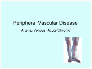
Peripheral Vascular syste m examination. Miss Sandy Abeysiri Clinical Teaching Fellow Bart’s Health NHS Trust. ‘Peripheral Vascular disease’. Arteries Or Veins (or Lymphatics – not covered this time) Progressive and chronic Aorto -iliac or infrainguinal

E N D
Peripheral Vascular system examination Miss Sandy Abeysiri Clinical Teaching Fellow Bart’s Health NHS Trust
‘Peripheral Vascular disease’ • Arteries Or Veins (or Lymphatics – not covered this time) • Progressive and chronic • Aorto-iliac or infrainguinal • Arterial : Aneurysmal, or Occlusive / Ischaemic • Venous: DVT / Varicose veins / Chronic insufficiency (Lymphatic: Lymphoedema / lymphangitis)
Assessment • Anatomy • History / clinical presentation • Aetiology or Causes / Risk Factors • Examination – special tests
History / Clinical Presentation • Full History – • Claudication – Site? • Rest Pain !!!!! • Ulceration? Chronic Wounds / poor healing • Bleeding / itching / skin changes / swelling / cellulitis • Impotence • Skin / sensation changes
RISK FACTORS • SMOKING! (even a few cigarettes/day) • Diabetes (esp poorly controlled) • Dyslipidaemia • Hypertension • Family Hx (CVS/PVD/dyslipidaemias) • Age • Ethnicity • Obesity (Vasculitides – active inflammation, hyperhomocysteinaemias)
atheroGenesis and atherosclerosis • LDL ingestion by mono/macrophage in subendothelial space – fatty streaks • Smooth muscle cells migrate – form atheroma with cap • Stenosis of vessel – decreased blood flow – reduction in oxygenation and nutrition to tissues (chronic) • Worse when increased demand eg.exercise and therefore symptoms
Inspection • REMEMBER inspection can give you clues to causes also – • Insulin Pens, Tar Staining • General inspection including hands and face Look closer at legs – • Skin changes / Hair changes / Colour changes • Ulcers / Wounds • Scars
Palpation • SUPINE – expose abdomen and legs: underwear left on initially, have a blanket for comfort Feel – Start distally – temperature / scars (compare L Vs R) Tibial shaft for oedema (pitting or non) – note upper level (severity) scrotum /abdominal wall maybe involved – unilateral disease – DVT or compression of large veins by node/tumour Non pitting – lymphatic diseases / hypothyroidism
Palpation(and auscultation) • Feel PULSES (against som’ hard) (and auscultate each for bruits at same time or you will forget!!!) DP / Post Tib / Pop / Femoral Abdo– MUST complete exam by assessing Abdominal Aorta, bruits and assess for sacral oedema… AT LEAST SAY- full abdo exam: ascites (severe CF), tender liver- capsule/veins, splenomegaly, full CVS exam
PULSES POPLITEAL Relax the fossa Use both hands to feel behind fat pad – almost like bimanual palp DEMO
Buerger’s test “Any hip pain if I lift your leg up straight…?” Angles ? 15-30 ischaemia - <20 severe
SPECIAL TESTS - ABPI • Ratio BP lower limb to BP Upper limb • Indicates - Severity of PVD • BP ankle – systolic DP or post Tib • BP arm – highest of left or right Brachial systolic • ABPI = BP ankle / BP arm
ABPI – interpretation • >1.2 – suggests calcification of vessels (age) • 1.2-1.0 – Normal Range • 0.9 – 1.0 – Acceptable (borderline abnormal) No referral needed • 0.8-0.9 – mild disease (manage risk factors) • 0.5 – 0.8 - moderate disease (routine referral) (mixed ulcers – bandage with care) • <0.5 – severe disease (URGENT referral) (no compression bandaging!!!)
Remember – acute ischaemia • Painful • Pale • Pulseless • ‘Perishingly’ cold • Paraesthetic • ?paralysed
Venous system • Position – this time STANDING • Remainder the same – Inspect / Palpate/ • Special tests • WEAR GLOVES!
Examination Inspect WHOLE leg – long saph / short saph regions If unilateral swelling – you may be asked to measure circumference (Use bony landmark as point of reference) Palpate – hard veins = Thrombosis, Tenderness = phlebitis Cough Impulse test: saphenofemoral valve (thrill – incompetence) Trendelenburg – incompetence of saphenofemoral valve (POSITIVE) If veins still fill up – incompetence lower - Perthes Test
Perthes test Same as Trendelenburg – but on standing release small vol blood into veins Ask patient to pump calves (stand up and down on tip toes) Veins become less tense if perforators have competent valves More tense if incompetence Note Pt will feel pain when veins fill up!!!! So BE VIGILANT and ready to STOP IMMEDIATELY PAIN TO PATIENT IN EXAM = BAD!!!!
DVT • Difficult clinical diagnosis • High index of suspicion!! • Unilateral swelling – and high risk (history) • Can possibly demonstrate difference in calf size • Differential diagnoses • When presenting include investigations • Wells Score