Download
1 / 25
300 likes | 700 Views
Biosensor in Medical Application. Reporter: AGNES Purwidyantri Student ID no: D0228005 Biomedical Engineering Dept. Classical definition of biosensor. Current Definition for Biosensors:.

E N D
Biosensor in Medical Application Reporter: AGNESPurwidyantri Student ID no: D0228005 Biomedical Engineering Dept.
Current Definition for Biosensors: A sensor that integrates a biological elementwith a physiochemical transducer to produce an electronic signal proportional to a single analyte which is then conveyed to a detector. www.imec.be/ovinter/static_research/BioHome.shtml
D. Grieshaber, R. MacKenzie, J. Voeroes and E. Reimhult, Sensors, 2008, 8, 1400–1458
Types of sensors • Physical sensor physical quantity: temperature, pressure, weight or force • Chemical sensor chemicals including ions • Biosensors biomolecules usually through chemical or biological interaction • Biomedical sensors: analytes relative to disease, healthcare or environmental protection. Ex: gases: CO2, O2, NH3. Hormones, glucose, etc
Biosensor classification based on FDA • Computer-related technology • Molecular medicine • Home and self‐care products • Minimally invasive procedures • Combination device/drug products • Organ substitute and assist devices
Basic Sensor Performance • Sensitivity • Linearity • Application range (Low/High limitation) • Response time • Lifetime • Reproducibility • Interference (selectivity)
Biosensor 1STComponent: Sensing Element The component used to bind the target molecule. Must be highly specific, stable under storage conditions, and immobilized. • DNA : ssDNA to complementary DNA • • Enzymes: with specificity and catalysis • • Antibody/antigen: immuno‐based, highly specific and strong binding • • Receptors/analytes : high specificity • • Chemical/chemical : chemical reactions • • Gas/solid materials : (metal, oxides, polymers… etc.) http://www.chemistry.wustl.edu/~edudev/LabTutorials/HIV/DrugStrategies.html
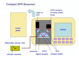
2ND Component: Transducer Acts as an interface, measuring the physical change that occurs with the reaction at the bioreceptor then transforming that energy into measurable electrical output. • Electrochemical sensors: Potentiometric& voltammetric (amperometric) • •Semiconductor‐based sensors: FETs, Schottky diodes • • Piezoelectric sensors: QCM, SAW • • Optical sensors: surface plasmon resonance (SPR), surface‐enhanced Raman scattering.
3RD Component: Detector Signals from the transducer are passed to a microprocessor where they are amplified and analyzed. The data is then converted to concentration units and transferred to a display or/and data storage device. www.modernmike.com
Static Characteristics • Accuracy • Resolution • Reproducibility • Statistical control • Static sensitivity • - the ratio of the incremental output quantity to the incremental • input quantity. • - zero drift, sensitivity drift • • Linearity • • Input ranges • • Input impedance
Biosensors for biotoxins and biologicalpathogens This scheme demonstrates the application of the infection process to ricin detection, since ricin specifically recognizes cell surface oligosaccharides, and the sugar based nanomaterials (A) were designed for use in surface plasmon resonance (B) and (C), and in a colloidal gold colorimetric assay for the qualitative detection of the toxin (D) Uzawa et al., Biosens. Bioelectron., 2008, 24, 923–927
Luo et al., Biosens. Bioelectron., 2010, 26, 1612–1617 (A) Schematic showing the biosensor structure and membrane assembly consisting of the application pad, adsorption pad, and the capture pad. (B) Detection scheme of the biosensor based on antibodies and magnetic separation of the analyte from mixed samples
DNA magnetic-based sensor Zhang et al., Biosens. Bioelectron., 2009 Schematic of the bio-barcode assay with (A) formation of the magnetic nanoparticles (MNPs) with the DNA probe complex including the target DNA and the barcode DNA on gold nanoparticles (Au-NPs) and (B) the separation of the complex and release of the barcode DNA
Biosensor on food quality test Li et al., Biosens. Bioelectron., 2010, 26, 1313–1319 Scheme of the process used to directly detect Salmonella on tomato surfaces using magnetoelastic biosensors
Medical biosensorsImmunology and infectious disease diagnosis Ma et al., Nat. Med., 2011, 17, 738–743. Layout of the single-cell barcode chip (SCBC) system. (a and b) Optical image of an SCBC system and cells isolated in microchambers; (c) scheme of cytokine detection by antibody barcode array; (d) scanned antibody barcode array; (e) calibration curve for recombinant cytokine proteins.
Cancer Diagnostic and monitoring – circulating tumor cell Cho et al.,ACSNano, 2012, 6, 7607–7614 (A) In the absence of target molecules, VEGF165, unfolded VEGF165 aptamer is electrostatically bound to a positively charged PLL-coated gold nanoparticle (GNP) surface and surface-enhanced fluorescence (SEF) of Cy3B conjugated with the VEGF165 aptamer is created by both a metal interaction increasing the radiative fluorescent decay rate of Cy3B and the local surface plasmon resonance (LSPR) enhancing the intensity of an incident light. (B) The interaction of the VEGF165 aptamer with its target induces the reversible conformation change of the aptamer and, consequently, the decreased electrostatic binding force. (C) As a result, the target-binding interaction of the aptamer causes the irreversible detachment of the aptamer from the GNP surface and avoids the SEF effect of Cy3B
Application in neuroscience Hai et al., Nat. Methods, 2010, 7 200–202. A mushroom-shaped microelectrode (gMmE) engulfed by a neuron for electrical coupling. (a and b) Schematic illustration of a neuron on a gold-spine electrode (a) and a flat electrode (b); (c) electron micrograph image of the cross section of a cell which engulfs three gold spine electrodes; (d) transmission electron micrograph of a gMmE engulfed by a cell; (e) three neurons cultured on gold spine electrode arrays; (f) action potential signal monitored from 8 spine electrodes.