Download
1 / 75

E N D
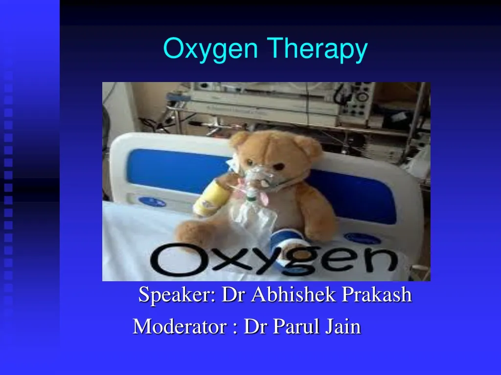
Oxygen Therapy Speaker: Dr AbhishekPrakash Moderator : Dr Parul Jain
HISTORY 1.Oxygen was discovered by Carl Wilhelm Scheele in Uppsala in 1773 but Priestly is often given priority because his work was published first. 2.Oxygen was also discovered by Priestly in 1774 who first realised its importance as a normal constituent of air and called it Dephlosgisticated Nitrous Acid.. 3.In 1777,Lavoisier named it oxygen. 4.Modern oxygen therapy initiated in 1917 by J.S.Haldane
Partial Pr of O2 in insp. gas (Pi o2) Oxygen Therapy Conc. of O2 (Fi o2) (Orthobaric) Total Pressure (Hyperbaric)
HYPERBARIC OXYGEN THERAPY It works on Henry’s Law which states that amount of gas dissolved in a liquid is directly proportional to its partial pressure. So,thePRESSURE GRADIENT is greatly increased between the arterial and hypoxic tissue and this allows an increasd rate of oxygen transport from blood to cells.Thus,Hyperbaric Oxygen therapy is EFFICIENT AND RAPID method of restoring cellular oxygenation. When a patient is given 100% oxygen under pressure, hemoglobin is saturated, but the blood can be hyperoxygenated by dissolving oxygen within the plasma.
HYPERBARIC PHYSICS AND PHYSIOLOGY The physics behind hyperbaric oxygen therapy (HBOT) lies within the ideal gas laws. The application of Boyle’s law (p1 v1 = p2 v2) is seen in many aspects of HBOT. This can be useful with embolic phenomena such as decompression sickness (DCS) or arterial gas emboli (AGE). As the pressure is increased, the volume of the concerning bubble decreases. This also becomes important with chamber decompression; if a patient holds her breath, the volume of the gas trapped in the lungs overexpands and causes a pneumothorax. Charles’ law ([p1 v1]/T1 = [p2 v2]/T2) explains the temperature increase when the vessel is pressurized and the decrease in temperature with depressurization. This is important to remember when treating children or patients who are very sick or are intubated. Henry’s law states that the amount of gas dissolved in a liquid is equal to the partial pressure of the gas exerted on the surface of the liquid. By increasing the atmospheric pressure in the chamber, more oxygen can be dissolved into the plasma than would be seen at surface pressure.
Application of HENRY’S LAW in Hyperbaric Oxygen therapy 0.003ml / 100ml of blood / mm PaO2 In normal state,100 ml of blood will dissolve 0.3 ml of oxygen only as it has a PaO2 of 100 mm of Hg.. 3 SCENARIOS 1.Breathing Air (PaO2 100mm Hg) 0.3ml / 100ml of blood 2.Breathing 100% O2 (PaO2 600mm Hg) 1.8ml / 100ml of blood 3.Breathing 100% O2 at 3 Atm. Pressure 5.4ml / 100ml of blood
HBOT AS HYPEROXYGENATION Most oxygen carried in the blood is bound to hemoglobin, which is 97% saturated at standard pressure. Some oxygen, however, is carried in solution, and this portion is increased under hyperbaric conditions due to Henry's law. Tissues at rest extract 5-6 mL of oxygen per deciliter of blood, assuming normal perfusion. Administering 100% oxygen at normobaric pressure increases the amount of oxygen dissolved in the blood to 1.5 mL/dL; at 3 atmospheres, the dissolved-oxygen content is approximately 6 mL/dL, which is more than enough to meet resting cellular requirements without any contribution from hemoglobin. Because the oxygen is in solution, it can reach areas where red blood cells may not be able to pass and can also provide tissue oxygenation in the setting of impaired hemoglobin concentration or function.
HBOT AND BACTERIAL KILLING HBOT increases the generation of oxygen free radicals, which oxidize proteins and membrane lipids, damage DNA, and inhibit bacterial metabolic functions. HBO is particularly effective against anaerobes and facilitates the oxygen-dependent peroxidase system by which leukocytes kill bacteria.
HBOT & VASOCONSTRICTION Hyperoxia in normal tissues causes vasoconstriction, but this is compensated by increased plasma oxygen content(almost 6ml%) and microvascular blood flow. This vasoconstrictive effect does, however, reduce posttraumatic tissue edema, which contributes to the treatment of crush injuries, compartment syndromes, and burns.
MODE OF ADMINISTRATION Oxygen at high pressure can be given from a pressure chamber.The patient then receives oxygen from from an ordinary mask and cylinder.A pressure of 2 atm is generally employed. 2 types of chamber are available: 1.Monoplace/Single Occupant A monoplace chamber compresses one person at a time, usually in a reclining position . The gas used to pressurize the vessel is usually 100% oxygen. Some chambers have masks available to provide an alternate breathing gas (such as air). Employees tend to the patient from outside of the chamber and equipment remains outside the chamber; only certain intravenous lines and ventilation ducts penetrate through the hull. 2.Multiplace HYPERBARIC OXYGEN BED: Rate of compression and decompression is controlled from an adjacent console
OXYGEN TRANSPORT Hemoglobin is transported as: 1.COMBINATION WITH HEMOGLOBIN About 98.5% of the oxygen in a healthy human being breathing air at sea level pressure is chemically combined with hemoglobin. 2 AS DISSOLVED IN PLASMA Around 1.5% which can be increased by increasing PaO2 by hyperbaric oxygen therapy(HENRY LAW)
Transport Contd.. Hemoglobin in blood leaving the lungs is about 98–99% saturated with oxygen, achieving an oxygen delivery of between 950 - 1150 mL/min[15] to the body. In a healthy adult at rest, oxygen consumption is approximately 200 - 250 mL/min,[15] and deoxygenated blood returning to the lungs is still approximately 75%[16][17] (70 to 78%)[15]saturated. Increased oxygen consumption during sustained exercise reduces the oxygen saturation of venous blood, which can reach less than 15% in a trained athlete; although breathing rate and blood flow increase to compensate, oxygen saturation in arterial blood can drop to 95% or less under these conditions
Oxygen Flux and Requirements The supply of oxygen is dependent upon the hemoglobin (Hb), O2 saturation % (SaO2) and cardiac output (Q). "Oxygen flux" denotes the total amount of oxygen delivered to the body per minute and is given by the equation: Oxygen flux= 1.34 x Hb in g/dL x (SaO2/100) x (Q in mL/min)/100 = 1000 mL/min
O2 Cascade :The Partial pressure of oxygen drops through various stages from 159 mm of Hg to low levels as 8-10 mm of Hg at mitochondria level….. Air Mitochondria..If Po2 falls below 1-2 m of Hg at mitochondrial level, AEROBIC METABOLISM stops ..which is known as PASTEUR POINT..
O2 Cascade 159mm Hg (20.95 % of 760) Atm. Air (dry) Humidification 6 Vol % (47mm Hg) Lower Resp. Tract (moist 37oc) 149mm Hg 20.95 % of 713 (760-47)
O2 Cascade Lower Resp. Tract (moist 37oc) 149mm Hg (20.95 % of 713) O2 consumption Alv. ventilation 101mm Hg (14 % of 713) or (15 % of 673) 673 = 760 – 47 – 40 Alveolar air PA O2 = FI O2 (Pb – 47) – PaCo2 x F = PI O2 –PaCo2 = PI O2 – PaCo2 if breathing 100% O2 R.Q
O2 Cascade 101mm Hg (14 % of 713) or (15 % of 673) 673 = 760 – 47 – 40 Alveolar air Venous admixture Arterial blood 97mm Hg Pa O2 = 100 – 0.3 x age (years) mm Hg A – a = 4 – 25 mmHg
O2 Cascade Pa O2 = 97mm Hg (Sat. > 95 %) Arterial blood Utilization by tissue Cell Mitochondria PO2 7 – 37 mmHg Mixed Venous blood PV O2 = 40mm Hg Sat. 75% Pasteur point – The critical level for aerobic metab. to continue (1 – 2 mmHg PO2 in mitochondria)
INDICATIONS OF OXYGEN THERAPY PULMONARY NON PULMONARY 1.Resuscitation(CPR) 2.Major Trauma 3.Major hemorrhage 4.Anaphylaxis 5.Acute Myocardial Infarction 6.Active Convulsions 7.Hypermetabolic states-Thyrotoxicosis,Hyperthermia,Anaemia 8.ANY ILLNESS CAUSING HYPOXEMIA 1.Acute Asthma 2.Acute Exacerbation of COPD-PaO2 ≤ 55mmHg or SaO2 ≤ 88% 3.Continuosly in COPD patients 4.Breathlessness in setting of END STAGE Cardiac or respiratory failures
HYPOXEMIA Criteria1.Documented hypoxemiaIn adults, children, and infants older than 28 days, arterial oxygen tension (PaO2) of < 60 mmHg or arterial oxygen saturation (SaO2) of < 90% in subjects breathing room air or with PaO2 and/or SaO2 below desirable range for specific clinical situationIn neonates, PaO2 < 50 mmHg and/or SaO2 < 88% or capillary oxygen tension (PcO2) < 40 mmHg
Hypoxia Vs Hypoxemia • Hypoxia-This is inadequate O2 tensions at cellular level and cannot be measured • Hypoxemia- This is defined as relative deficiency of O2 in arterial blood.
Types of hypoxia 1. Hypoxic hypoxia ( decrease diffusion of O2 across the alveolar-capillary membrane -low inspired FiO2 -V/Q inequalities -increased shunt(eg cardiac anomalies) 2. Stagnant hypoxia (decreased cardiac output resulting in increased systemic transit time -Shock -Vasoconstrictio 3. Anaemic hypoxia ( decreased O2 carrying capacity in the blood) -Anaemia Histotoxic Hypoxia(inability to utilise available oxygen) -cyanide poisoning
Benefit of O2 therapy in Hypoxia Hypoxic hypoxia (gas phase) + + + Anaemic hypoxia (fluid phase – const.) + Stagnant hypoxia (fluid phase – flow) + Histotoxic hypoxia (tissue phase) -
General Goals/Objectives 1.Correcting Hypoxemia 2.Decreasing Symptoms of Hypoxemia Lessen Dyspnoea/work of breathing Improve Mental function 3.Minimising Cardiopulmonary Workload
Goals CONTD.. 3.Minimising Cardiopulmonary Workload Cardiopulmonary system would compensate for HYPOXEMIA by: -Increasing ventilation to get more oxygen in the lungs and in the blood leading to INCREASED WORK OF BREATHING. -Increasing Cardiac output to get more oxygenated blood to tisses which puts EXTRA LOAD ON HEART,IF DISEASED. -HYPOXIA causes pulmonary vasoconstriction and Pulm hypertension which causes increased workload on right side of heart.
Oxygen therapy To ensure safe and effective treatment remember: • Oxygen is a prescription drug. • Prescriptions should include – • Flow rate. • Delivery system. • Duration. • Instructions for monitoring.
Oxygen therapy Oxygen delivery methods: • All systems require. • Oxygen supply. • Flow meter. • Oxygen tubing. • Delivery device. • (Humidifier).
Normal Anatomic Reservoir (50ml) 3 Ltr/min = 50 ml/Sec No capacity system sSmall capacity system 100-200 ml Large capacity System 1l-2L
Delivery Systems: CONCEPT OF ANATOMICAL RESERVOIR: This is air contained in Oropharynx and Nasopharynx which is about 1/3rdof anatomical dead space or 50 ml.. Low flow systems with no capacity systems-NASAL CATHETERS and NASAL CANNULA use it as a reservoir which empties into lungs with each inspiration even when the mouth is wide open..
Oxygen therapy Humidification • Is recommended if more than 4 litres/min is delivered. • Helps prevent drying of mucous membranes. • Helps prevent the formation of tenacious sputum.
HIGH FLOW CLASSIFICATION AIR ENTRAINMENT SYSTEMS BLENDING DEVICES 1.MANUAL GAS MIXER 2.OXYGEN BLENDER • 1.AE MASK(VENTURI MASK) • 2.AE NEBULISERS MECHANICAL VENTILATION using ventilator and ETT is a High Flow System..
NO CAPACITY SYSTEMS NASAL CATHETER NASAL CANNULA/NASAL PRONGS 2 PRONGS protrude 1 cm into nares and other end is attached to oxygen source FiO2 is more unpredictable than with nasal catheter. Humidification becomes an important part in higher flow rates(>4 L/min).. • Consists of a soft tube 8-14 FG size with several holes at its end.. • Length should be from angle of mouth to tragus • Its inserted through nostrils into oropharynx, just below soft palate.. • It should be changed to other nostril every 8-10 hours
NO CAPACITY SYSTEMS NASAL CATHETER NASAL CANNULA/NASAL PRONGS ADVANTAGES: Allows continuous flow of oxygen during routine nursing when patient is eating or oral suction is done. Can be used when the nasogastric tube is occuding one nostril. DISADVANTAGES: Higher flow rates (>4L/min) may dry the nasal mucosa and produce local irritation anddermatitis.So,HUMIDIFICATION of oxygen is essential. ADVANTAGES: Longer the end expiratory pause, higher the FiO2 FiO2 delivered ranges from 25-40% Roughly,Fio2 changes by 4% for every L/min change in oxygen flow rate DISADVANTAGE: Causes greater irritation of nasal mucosa Gastric dilatation with high flows
FACE MASKS 1.SIMPLE FACEMASK Around 100-200 ml of air gathers in this mask. Air enters through exhalation ports and around face mask. Oxygen Flow rate(L/min) FiO2 5-6 0.40 6-7 0.50 7-8 0.60 It has vents/exhalation ports on the sides for the room air to leak in and thereby diluting the source oxygen. Also allows exhaled Co2 to escape. Used when oxygen delivery is required for short periods<12 hours
Simple Face Mask Thus,simple Face mask delivers the oxygen concentration from 40%-60% at a flow rate of 5L to 8L/min respectively. CAUTION: Due to risk of retaining/rebreathing CO2,we should never apply a simple face mask with a delivery rate of less than 5 L/min.
FACE MASKS 2.PARTIAL REBREATHER MASK Utilises 1litre reservoir bag and mask. Delivers oxygen concentrations of 60-90% at a flow rate of 6L to 8L/min respectively. ONE VALVE 1st third(dead space) is breathed into reservoir bag and rebreathed. air enters throug exhalation ports and around the mask(AS IN SIMPLE FACE MASK).
FACE MASKS Contd. 3.NON REBREATHER MASK • Utilises ONE WAY VALVES-2 VALVES • Between reservoir and bag • On one exhalation port.Note that other port is same as in simple face mask... • It can deliver highest possible oxygen concentration(95% to 100%) at flow rates of 10 to15L/min,provided leak free system is provided,which is rare.Hence,>70% FiO2 is rare • One way valves prevent room and expired air from diluting the oxygen concentration. • Reservoir bag must be seen to expand freely.
Variables O2 flow rate Patient factors • Inspiratory flow rate • Expiratory time (active exp. flow + exp. pause) Device factors • Physical volume (capacity) • Vent resistance (tight fit)
High Flow devices Supplies given FiO2@ flows higher than inspiratory demand. These use Air Entrainment(AE) systems or Blenders. AE devices are- 1.AEM(Ventimask) 2.AE Nebuliser(Large volume nebuliser) Peak Inspiratory Flow is 3 times minute ventilation.Since 20L/min is upper limit of minute ventilation,maximuminspiratory flow of 60L/min is possible with these devices..