Download
1 / 56
570 likes | 1.06k Views
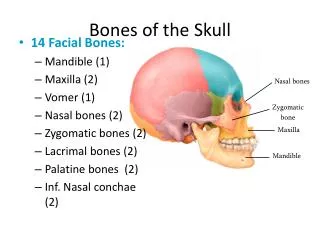
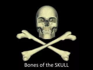
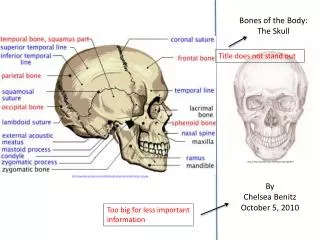
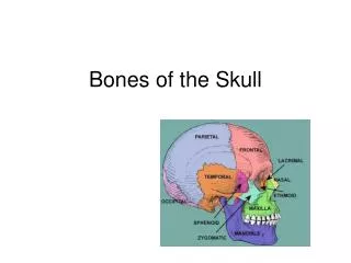
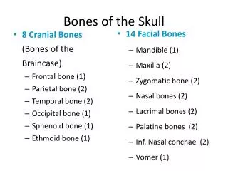
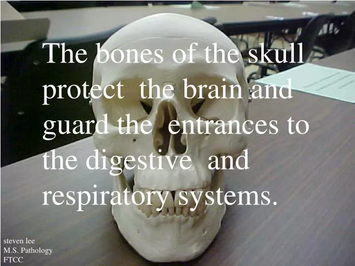
The bones of the skull protect the brain and guard the entrances to the digestive and respiratory systems. steven lee M.S. Pathology FTCC. steven lee M.S. Pathology FTCC. Frontal bone. Zygomatic bone. maxilla. vomer. steven lee M.S. Pathology FTCC. mandible. Frontal bone.

E N D
The bones of the skull protect the brain and guard the entrances to the digestive and respiratory systems. steven lee M.S. Pathology FTCC
steven lee M.S. Pathology FTCC
Frontal bone Zygomatic bone maxilla vomer steven lee M.S. Pathology FTCC mandible
Frontal bone Supraorbital Foramen (frontal bone) Glabella (frontal bone) Zygomatic Process (frontal bone) Supraorbital Margin (frontal bone) Infraorbital foramen (maxillary bone) Middle nasal concha (ethmoid bone) Inferior nasal concha maxilla Perpendicular plate (ethmoid bone) Alveoli processes (maxillary bone) vomer Alveoli processes (mandible) steven lee M.S. Pathology FTCC mandible
Frontal eminence Superciliary arch (frontal bone) steven lee M.S. Pathology FTCC
steven lee M.S. Pathology FTCC
Middle nasal concha (ethmoid bone) Perpendicular plate (ethmoid bone) Inferior nasal concha Infraorbital foramen (maxillary bone) steven lee M.S. Pathology FTCC vomer
steven lee M.S. Pathology FTCC
Optic foramen or canal (sphenoid bone) Superior orbital fissure (sphenoid bone) Inferior orbital fissure (maxillary bone) steven lee M.S. Pathology FTCC
When the supraorbital foramen is not completely enclosed by bone, there is a supraorbital notch, rather than a foramen, on the orbital rim, as is the case for this specimen. steven lee M.S. Pathology FTCC
Optic canals permit passage of the optic nerves from the eyes to the brain. steven lee M.S. Pathology FTCC
The inferior orbital fissure permits passage of cranial nerves and blood vessels. steven lee M.S. Pathology FTCC
The superior orbital fissure permits passage of cranial nerves and blood vessels to the orbit steven lee M.S. Pathology FTCC
steven lee M.S. Pathology FTCC
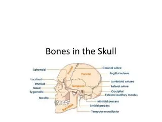
Frontal bone Parietal bone lacrimal nasal Temporal bone Occipital bone Zygomatic bone maxilla mandible steven lee M.S. Pathology FTCC
steven lee M.S. Pathology FTCC Zygomatic process (frontal bone) Frontal bone Coronal suture Parietal bone Frontal process (zygomatic bone) Zygomatic process (Temporal bone) lacrimal nasal Condylar process (mandible) Occipital bone Temporal process Zygomatic bone External auditory meatus (temporal bone) maxilla ramus Coronoid process (mandible) External occipital protuberance angle Mastoid process (temporal bone) mandible Styloid process (temporal bone)
steven lee M.S. Pathology FTCC
Supra meatal triangle Mandibular condyle Post glenoid process External auditory meatus (temporal bone) Tympanic part mandible Mastoid process (temporal bone) steven lee M.S. Pathology FTCC Styloid process (temporal bone)
steven lee M.S. Pathology FTCC
Frontal bone Zygomatic process (frontal bone) Sphenoid bone Temporal bone Frontal process (zygomatic bone) Lacrimal bone Nasal bone Ethmoid bone Zygomatic arch Zygomatic process (temporal bone) Temporal process (zygomatic bone) Zygomatic bone Condylar process (mandible) Coronoid process (mandible) Mandibular notch steven lee M.S. Pathology FTCC maxilla mandible
steven lee M.S. Pathology FTCC
Sagittal suture Parietal bone Lambdoidal suture Parietal bone Occipital bone Superior nuchal line External occipital protuberance Inferior nuchal line Middle nuchal line Condyloid canal Mastoid process (temporal bone) steven lee M.S. Pathology FTCC Styloid process (temporal bone)
steven lee M.S. Pathology FTCC
Frontal bone Coronal suture Parietal bone Sagittal suture Parietal bone steven lee M.S. Pathology FTCC Occipital bone
steven lee M.S. Pathology FTCC
Greater wing (sphenoid bone) Lesser wing (sphenoid bone) Jugular foramen Foramen magnum surrounds the connection between the brain and spinal cord. Mastoid foramen Hypoglossal canals steven lee M.S. Pathology FTCC
Location of foramen rotundum (sphenoid bone), although not shown on this model Foramen lacerum (sphenoid bone) Lesser wing (sphenoid bone) Foramen ovale (sphenoid bone) Greater wing (sphenoid bone) Foramen spinosum (sphenoid bone) Petrous portion (temporal bone) Petrous portion (temporal bone) Foramen magnum Internal occipital crest Internal occipital protuberance steven lee M.S. Pathology FTCC
steven lee M.S. Pathology FTCC
Cribiform plate (ethmoid bone) Crista galli (ethmoid bone) Lesser wing (sphenoid bone) Sulcus chiasmatis Tuberculum sellae Sella turcica (sphenoid bone) Fossa hypophysis Dorsum sellae steven lee M.S. Pathology FTCC
Crista galli (ethmoid bone) The falx cerebri, a membrane that stabilizes the position of the brain, attaches to this ridge. Lesser wing (sphenoid bone) Greater wing (sphenoid bone) Sella turcica (sphenoid bone) steven lee M.S. Pathology FTCC
steven lee M.S. Pathology FTCC
Optic canal (sphenoid bone) The hypophyseal fossa, the depression within the sella turcica houses and protects the pituitary gland. steven lee M.S. Pathology FTCC
steven lee M.S. Pathology FTCC
Lesser wing (sphenoid bone) Petrous portion (temporal bone) Lacrimal canal (lacrimal bone) nasal Alveoli processes (maxillary bone) Internal acoustic canal (temporal bone) Squamous portion (temporal bone) mandible ramus body steven lee M.S. Pathology FTCC angle Mastoid process (temporal bone)
steven lee M.S. Pathology FTCC
Petrous portion (temporal bone) Internal auditory meatus (temporal bone) The internal auditory canal (meatus) carries blood vessels and nerves to the inner ear. steven lee M.S. Pathology FTCC
steven lee M.S. Pathology FTCC
Palatine bones mandible Zygomatic arch Palatine process (maxilla) Foramen magnum Occipital condyles steven lee M.S. Pathology FTCC External occipital ptotuberance
Occipital condyles are the site of articulation between the skull and the 1st cervical vertebrae (atlas). Occipital condyles steven lee M.S. Pathology FTCC
steven lee M.S. Pathology FTCC
mandible Palatine process (maxillary bone) Palatine bone steven lee M.S. Pathology FTCC Palatine bone
steven lee M.S. Pathology FTCC
Pterygoid processes medial medial lateral lateral Pterygoid plates (sphenoid bone) vomer steven lee M.S. Pathology FTCC Foramen magnum
steven lee M.S. Pathology FTCC
steven lee M.S. Pathology FTCC maxilla Coronoid process Maxillary alveoli processes Mandibular notch Condylar process ramus Oblique line Mental foramen Mandibular alveoli processes body Mandibular angle mandible Mental protuberance
steven lee M.S. Pathology FTCC temporalis buccinator masseter mentalis
steven lee M.S. Pathology FTCC
Maxillary bone Alveoli processes Alveoli processes steven lee M.S. Pathology FTCC mandible
FETAL SKULL steven lee M.S. Pathology FTCC
Metopic suture At birth, there are 2 frontal bones that articulate along the metopic suture. Although the suture generally disappears by age 8 as the bones fuse, the adult skull commonly retains traces of the suture line. steven lee M.S. Pathology FTCC