Download
1 / 50
500 likes | 634 Views
Chapter 21: The Thigh, Hip, Groin, and Pelvis. Jennifer Doherty-Restrepo, MS, LAT, ATC Academic Program Director, Entry-Level ATEP Florida International University Acute Care and Injury Prevention. Anatomy of the Thigh. Review. Quadriceps. Insertion at proximal patella via common tendon

E N D
Chapter 21: The Thigh, Hip, Groin, and Pelvis Jennifer Doherty-Restrepo, MS, LAT, ATC Academic Program Director, Entry-Level ATEP Florida International University Acute Care and Injury Prevention
Anatomy of the Thigh Review
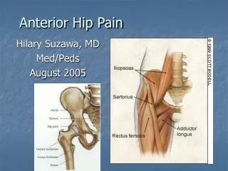
Quadriceps • Insertion at proximal patella via common tendon • Pre-patellar tendon • Rectus femoris = bi-articulate muscle • Only quad muscle that also crosses the hip • Extends knee and flexes the hip • Important: distinguish between knee extensors and hip flexors • Injury evaluation • Treatment and rehabilitation programs
Hamstrings • Cross the knee joint posteriorly • All hamstrings, except the short of head of the biceps femoris, are bi-articulate • Crosses the hip joint as well • Forces dependent upon position of both knee and hip • Important: distinguish between knee flexors and hip extensors • Injury evaluation • Treatment and rehabilitation programs
Thigh Injuries: Quadriceps Contusions • Etiology • MOI = severe impact, direct blow • Extent (depth) of injury depends upon… • Force • Degree of thigh relaxation • Signs and Symptoms • Pain, transitory loss of function, immediate effusion (palpable) • Graded 1 - 4 = superficial to deep • Increased loss of function 1 - 4 • Decreased ROM 1 - 4 • Decreased strength 1 - 4
Thigh Injuries: Quadriceps Contusions • Management • RICE • NSAID’s and analgesics • Crutches, if indicated • Aspiration of hematoma • Ice post exercise or re-injury • Follow-up care • ROM exercises • PRE in pain-free ROM • Modalities • Heat • Massage • Ultrasound to prevent myositis ossificans
Thigh Injuries: Myositis Ossificans Traumatica • Etiology • Formation of ectopic bone • MOI = repeated blunt trauma • May be the result of improper thigh contusion treatment (too aggressive) • Signs and Symptoms • X-ray shows Ca++ deposit 2 - 6 weeks post injury • Pain, weakness, swelling, tissue tension, point tenderness, and decreased ROM • Management • Treatment must be conservative • May require surgical removal
Thigh Injuries: Quadriceps Muscle Strain • Etiology • MOI = over-stretching or too forceful contraction • Signs and Symptoms • Pain, point tenderness, spasm, loss of function, and ecchymosis • Superficial strain results in fewer S&S than deeper strain • Complete tear results in deformity • Athlete displays little disability and discomfort
Thigh Injuries: Quadriceps Muscle Strain • Management • RICE • NSAID’s and analgesics • Manage swelling • Compression, crutches • Stretching • PRE strengthening exercises • Neoprene sleeve for added support
Thigh Injuries: Hamstring Muscle Strains • Etiology: multiple theories of injury • Hamstrings and quadriceps contract together • Change from hip extender to knee flexor • Fatigue • Posture • Leg length discrepancy • Lack of flexibility • Strength imbalances
Signs and Symptoms Pain in muscle belly or point of attachment Capillary hemorrhage Ecchymosis Grade 1 Pain with movement Point tenderness <20% of fibers torn Grade 2 Partial tear <70% of fibers torn Sharp snap or tear Severe pain Loss of function Grade 3 Rupture of tendinous or muscular tissue >70% muscle fiber tearing Severe hemorrhage Disability Edema Loss of function Ecchymosis Palpable mass or gap Thigh Injuries: Hamstring Muscle Strains
Management RICE, NSAID’s and analgesics Modalities PRE exercises When soreness is eliminated, focus on eccentrics strengthening Recovery may require months to a full year Scaring increases risk of injury recurrence of Grade I Do not resume full activity until complete function restored Grade 2 and 3 Should treat conservatively Gradual return to stretching and strengthening in later stages of healing Thigh Injuries: Hamstring Muscle Strains
Thigh Injuries: Acute Femoral Fractures • Etiology • Fracture in middle third of femoral shaft • MOI = great deal of force • Signs and Symptoms • Pain, swelling, deformity, muscle guarding • Leg with fx positioned in hip adduction and ER • Leg with fx may appear shorter • Management • Medical emergency! • Treat for shock, splint, refer • Analgesics and ice
Thigh Injuries: Femoral Stress Fractures • Etiology • Overuse (10-25% of all stress fractures) • MOI = excessive downhill running or jumping • Often seen in endurance athletes • Signs and Symptoms • Persistent pain in thigh/groin region • X-ray or bone scan will reveal fracture • Positive Trendelenburg’s sign • Management • Prognosis will vary depending on location • Fx in shaft and medial to femoral neck heal well with conservative management • Fx lateral to femoral neck are more complicated
Functional Anatomy • Hip Joint • True ball and socket joint • Intrinsic stability • Moves in all three planes, particularly during gait • Pelvis • Moves in all three planes • Anterior tilting • Changes degree of lumbar lordosis • Lateral tilting • Changes degree of hip abduction
Assessment of the Hip and Pelvis • Injuries to the hip or pelvis cause major disability in the lower limbs, trunk, or both • Low back may also become involved • History • Onset (sudden or slow?) • Previous history? • Mechanism of injury? • Pain description, intensity, quality, duration, type, and location?
Assessment of the Hip and Pelvis • Observation • Symmetry - hips, pelvis tilt (anterior/posterior) • Lordosis or flat back • Lower limb alignment • Knees, patella, feet • Pelvic landmarks • ASIS, PSIS, iliac crest • Standing on one leg • Pubic symphysis pain or drop to one side • Ambulation
Special Tests: Leg Length Discrepancy • True or anatomical • Shortening may be equal throughout limb or localized in femur or lower leg • Measure from ASIS to medial malleolus • Apparent or functional • May result due to lateral pelvic tilt, flexion, or adduction deformity • Measure from umbilicus to medial malleolus
Hip and Groin Injuries Groin Strain • Etiology • Injury usually occurs to the adductor longus • MOI = running, jumping, or twisting with hip external rotation; over-stretching; or too forceful contraction • Signs and Symptoms • Sudden twinge or tearing during movement • Pain, weakness, and internal hemorrhaging
Hip and Groin Injuries Groin Strain (continued) • Management • RICE • NSAID’s and analgesics • Rest is critical • Modalities • Daily whirlpool and cryotherapy • Ultrasound • Delay exercise until pain free • Restore normal ROM and strength • Provide support with elastic wrap
Hip and Groin Injuries Trochanteric Bursitis • Etiology • Inflammation of bursa at greater trochanter • Insertion site for gluteus medius and where IT-band passes over the greater trochanter • Signs and Symptoms • Lateral hip pain that may radiate down the leg • Point tenderness over greater trochanter • IT-band and TFL tests should be performed
Hip and Groin Injuries Trochanteric Bursitis (continued • Management • RICE • NSAID’s and analgesics • ROM and PRE exercises for hip abductors and external rotators • Phonophoresis • Evaluate biomechanics and Q-angle • Runners should avoid inclined surfaces
Hip and Groin Injuries Sprains of the Hip Joint • Etiology • Unusual movement exceeding normal ROM • MOI = force from opponent/object, or, trunk forced over planted foot in opposite direction • Signs and Symptoms • Pain, which increases with hip rotation • Inability to circumduct hip • Similar S&S to stress fracture
Hip and Groin Injuries Sprains of the Hip Joint (continued) • Management • RICE • NSAID’s and analgesics • Depending on severity, crutches may be required • ROM and PRE are delayed until hip is pain-free • X-rays or MRI should be performed to rule out a possible fracture
Hip and Groin Injuries Dislocated Hip • Etiology • Result of traumatic force directed along the long axis of the femur • Posterior dislocation more common • Hip flexed, adducted, and internally rotated • Knee flexed • Rarely occurs in sport • Signs and Symptoms • Flexed, adducted, and internally rotated hip • Palpation reveals displaced femoral head • Medical emergency • Compications include soft tissue damage, neurological damage, and possible fracture
Hip and Groin Injuries Dislocated Hip (continued) • Management • Immediate medical care • Blood and nerve supply may be compromised • Contractures may further complicate reduction • 2 weeks immobilization • Crutch use for at least one month
Hip and Groin Injuries Avascular Necrosis • Etiology • Temporary or permanent loss of blood supply to the proximal femur • MOI = traumatic conditions (ie: hip dislocation) or non-traumatic conditions (ie: steroids, blood coagulation disorders) • Signs and Symptoms • Possibly no S&S in early stages • Develop over the course of months to a year • Joint pain with weight bearing, progressing to pain at rest • Limited ROM • Osteoarthritis may develop
Hip and Groin Injuries Avascular Necrosis (continued) • Management • Must be referred for X-ray, MRI, or CT scan • Most cases will ultimately require surgery • Conservative treatment • Non-weight bearing;ROM exercises; e-stim for bone growth; medication to treat pain • Limit necrosis • Reduce fatty substances, which react with corticosteroids • Limit blood clotting in the presence of clotting disorders
Hip Problems in the Young Athlete Legg Calve’-Perthes Disease (Coxa Plana) • Etiology • Avascular necrosis of the femoral head in child ages 4-10 • MOI = trauma (accounts for 25% of cases) • Signs and Symptoms • Pain in groin • Referred pain to the abdomen or knee • Limping • may exhibit limited ROM
Hip Problems in the Young Athlete Legg Calve’-Perthes Disease (continued) • Management • Bed rest to alleviate synovitis • Brace to avoid direct weight bearing • With early treatment, the femoral head may re-ossify and revascularize • Complications • If not treated early, will result in ill-shaping • May develop into osteoarthritis in later life
Hip Problems in the Young Athlete Slipped Capital Femoral Epiphysis • Etiology • Found mostly in tall boys between ages 10-17 • May be growth hormone related • MOI = trauma (accounts for 25% of cases) • 25% of cases are seen in both hips • Femoral head slippage on X-ray appears in posterior and inferior direction
Hip Problems in the Young Athlete Slipped Capital Femoral Epiphysis (continued) • Signs and Symptoms • Pain in groin that progresses over weeks or months • Hip and knee pain during passive and active motion • Limitations of hip abduction, flexion, and medial rotation • Limp • Management • Minor slippage • Rest and non-weight bearing may prevent further slippage • Major slippage results in displacement • Requires surgery • If condition goes undetected or if surgery fails, severe problems will result
Hip Problems in the Young Athlete The Snapping Hip Phenomenon • Etiology • Common in young female dancers, gymnasts, and hurdlers • MOI = repetitive movement that leads to muscle imbalance • Related to narrow pelvis, increased hip abduction, and limited lateral rotation • Hip stability is compromised
Hip Problems in the Young Athlete The Snapping Hip Phenomenon (continued) • Signs and Symptoms • Pain while balancing on one leg • Possible inflammation • Management • ROM exercises to increase flexibility • Flexion and lateral rotation • Cryotherapy and ultrasound may be utilized • PRE exercises to strengthen weak muscles
Pelvic Injuries Contusion (hip pointer) • Etiology • Contusion of iliac crest or abdominal musculature • MOI = direct blow • Signs and Symptoms • Pain, spasm, and transitory paralysis • Decreased ROM due to pain • Rotation of trunk, thigh/hip flexion
Pelvic Injuries Contusion (hip pointer) continued • Management • RICE for at least 48 hours • NSAID’s, • Bed rest 1 - 2 days • Referral must be made for X-ray • Modailities • Ice massage, ultrasound, occasionally steroid injection • Recovery lasts 1 - 3 weeks
Pelvic Injuries Stress Fractures • Etiology • Seen in distance runners – more common in women than men • MOI = repetitive cyclical forces from ground reaction forces • Common sites include inferior pubic ramus, femoral neck, and subtrochanteric area of the femur • Signs and Symptoms • Groin pain • Aching sensation in thigh that increases with activity and decreases with rest • Standing on one leg may be impossible • Deep palpation results in point tenderness
Pelvic Injuries Stress Fractures (continued) • Management • Rest for 2 - 5 months • Crutch walking • Especially for ischium and pubis stress fractures • X-rays are usually normal for 6 -10 weeks, therefore a bone scan will be required to detect the stress fracture • Swimming can be used to maintain CV fitness • Breast stroke should be avoided
Pelvic Injuries Avulsion Fractures and Apophysitis • Etiology • Common sites include ischial tuberosity, AIIS, and ASIS • MOI = sudden accelerations and decelerations • Signs and Symptoms • Sudden localized pain • Limited ROM • Pain, swelling, point tenderness • Muscle testing increases pain
Pelvic Injuries Avulsion Fractures and Apophysitis (continued) • Management • X-ray required for diagnosis • RICE, NSAID’s, crutch “toe-touch” walking • ROM exercises • PRE exercises • When 80 degrees of ROM have been regained • Return to play when full ROM and strength are restored