Download

1 / 13
160 likes | 1.18k Views
Diaphragm. Objectives Discuss the origin and insertion of diaphragm. Enlist the openings (with their contents) in the diaphragm. Give the motor and sensory nerve supply of the diaphragm. Diaphragm

E N D
Objectives Discuss the origin and insertion of diaphragm. Enlist the openings (with their contents) in the diaphragm. Give the motor and sensory nerve supply of the diaphragm
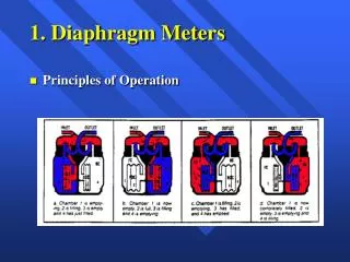
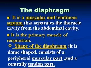
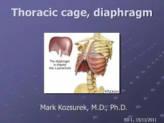
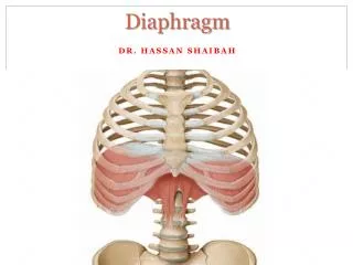
Diaphragm The diaphragm is a thin muscular and tendinous septum that separates thorax & abdominal cavities. It is pierced by the structures that pass between the chest and the abdomen. The diaphragm is the most important muscle of respiration. It is dome shaped and consists of a peripheral muscular part, which arises from the margins of the thoracic opening, and a centrally placed tendon. The origin of the diaphragm can be divided into three parts:
Origin of the diaphragm A sternal part arising from the posterior surface of the xiphoid process A costal part arising from the deep surfaces of the lower six ribs and their costal cartilages & forms the right & left domes A vertebral/lumbar part arising from upper three lumbar vertebrae; forms the right & left crura & the arcuate ligaments
Shape of the Diaphragm • It is studied as • Central tendon • Right & left crus • Right & left dome
Insertion of the Diaphragm The diaphragm is inserted into a central tendon. The superior surface of the tendon is partially fused with the inferior surface of the fibrous pericardium. Some of the muscle fibers of the right crus pass up to the left and surround the esophageal orifice in a slinglike loop. These fibers appear to act as a sphincter and possibly assist in the prevention of regurgitation of the stomach contents into the thoracic part of the esophagus.
Crura & Arcuate Ligaments The right crus arises from the sides of the bodies of the L 1-3 & IV discs; the left crus arises from the sides of the bodies of the L 1-2 & IV disc. Lateral to the crura the diaphragm arises from the medial & lateral arcuate ligament. The medial arcuate ligament extends from the side of the body of the second lumbar vertebra to the tip of the transverse process of the first lumbar vertebra. The lateral arcuate ligament extends from the tip of the transverse process of the first lumbar vertebra to the lower border of the 12th rib. The medial borders of the two crura are connected by a median arcuate ligament which crosses over the anterior surface of the aorta
Functions of the Diaphragm Muscle of inspiration: On contraction the diaphragm pulls its central tendon down and increases the vertical diameter of the thorax. The diaphragm is the most important muscle used in inspiration. Muscle of abdominal straining: The contraction of the diaphragm assists the contraction of the muscles of the anterior abdominal wall in raising the intra-abdominal pressure for micturition, defecation, and parturition. Weight lifting muscle: In a person taking a deep breath and holding it (fixing the diaphragm), the diaphragm assists the muscles of the anterior abdominal wall in raising the intra-abdominal pressure. Before doing this make sure that a person have adequate sphincteric control of the bladder and anal canal under these circumstances. Thoracoaabdominal pump: The descent of the diaphragm decreases the intrathoracic pressure & increases the intra-abdominal pressure. This compresses the blood in the inferior vena cava and forces it upward into the right atrium of the heart. Within the abdominal lymph vessels is also compressed, and its passage upward within the thoracic duct is aided by the negative intrathoracic pressure. The presence of valves within the thoracic duct prevents backflow.
Openings in the Diaphragm 8 10 12 The diaphragm has three main openings: The caval opening lies at the level of the T 8 vertebra in the central tendon. Inferior vena cava & branches of the right phrenic nerve. The esophageal opening lies at the level of the T 10 vertebra in a sling of muscle fibers derived from the right crus at the left of median plane. Esophagus, the right and left vagus nerves, the esophageal branches of the left gastric vessels, & the lymph vessels. The aortic opening lies anterior to the body of the T 12 vertebra between the crura. Aorta, thoracic duct, & azygos vein.
Nerve Supply of the Diaphragm Motor nerve supply: The right and left phrenic nerves (C3, 4, 5) Sensory nerve supply: The parietal pleura and peritoneum covering the central surfaces of the diaphragm are from the phrenic nerve and the periphery of the diaphragm is from the lower six intercostal nerves.
Blood supply • The phrenic artery. • The musculocutaneous. • Branches of the mammary artery. • The intercostals.