Download
1 / 24
240 likes | 370 Views
a 3-Dimensional Microarray Substrate. BioChip Ventures Division. What is a microarray?. Advantages multiplexing and miniaturization throughput parallel analysis sample volume reduction. target. probe. Protein Microarray Applications. * DNA - protein interaction

E N D
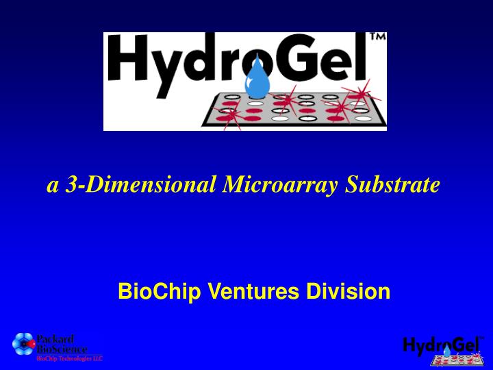
a 3-Dimensional Microarray Substrate • BioChip Ventures Division
What is a microarray? • Advantages • multiplexing and miniaturization • throughput • parallel analysis • sample volume reduction target probe
Protein Microarray Applications * DNA - protein interaction * Protein - protein interactions * Enzyme-substrate analysis * Protein profiling * Antibody characterization * Small molecule screening Image courtesy of Dr. Gavin MacBeath, Bauer Center for Genomics Research, Harvard University
Desirable Substrate Properties for Protein Microarray Applications • Protein compatible • High probe loading capacity • Low inherent fluorescence and nonspecific binding background • Consistent, uniform product • Ease of use
HydroGelTM Performance Validation • Printing compatibility • Inherent fluorescent background • Loading capacity of substrate • Protein compatibility • Nonspecific background • Multiplexed assay performance
Printing Compatibility Packard BioChip ArrayerTM (Piezo) Feature Size ~200 um Packard SpotArray 24 (Split Pin) Feature Size ~150 um HydroGelTM coated slides are compatible with both contact and non-contact printers (examples shown above). This was verified on two other types of commercially available and “home-made” instruments.
Texas Red CY3 CY5 FITC HydroGelTM coated slide 4.14 X 1.76 X 1.79 X 1.70 X Aldehyde glass 2.44 X 4.39 X 2.91 X 1.62 X Nitrocellulose coated slide 528 X 498 X 190 X 34 X Inherent Fluorescent Background Blank substrates were scanned on a ScanArrayTM 5000 microarray scanner in the channels indicated (laser power:100%, PMT gain: 75%).
Protein Loading Capacity IgG and Streptavidin were printed on HydroGelTM coated slides and aldehyde glass slides at the indicated concentrations to compare loading capacity.
Protein Penetration Demonstrated by Confocal Fluorescent Microscope Measurement ~70% penetration of a 160 kD protein starting ending 1.9 µm per section in Z axis
Print probes Immobilize and wash Incubate with target sample Wash and detect
Anti-bovine IgG Anti-avidin (negative control) HydroGelTM coated slide aldehyde glass nitrocellulose coated slide Non-Specific Background in a Direct Fluorescence Assay on Serum
Low Nonspecific Background HydroGelTM Coated Slide Poly-lysine based slide Targets:Cy3- and Cy5-labeled patient serum samples Images courtesy of Dr. Brian Haab (Van Andel Research Institute, Grand Rapids, MI).
Protein Compatibility * Calculated as the X value when Y is set to 2-fold the standard deviation of the background ** High inherent fluorescence of this substrate masks the signal generated by the two lowest enzyme concentrations.
ELISA: A Powerful Research Tool Representative commercial ELISA for IFN-g shows detection range of approximately 10-1000 pg/mL (2 log dynamic range)
Detection Complex For Sandwich Assays Texas Red conjugated Streptavidin Biotinylated detection antibody Target (cytokine) Capture antibody
IL-6 IFN-g IL-2 Det. Control IL-1b TNF-a IL-13 Neg. control replicates IL-6 IFN-g IL-2 Det. Control IL-1b TNF-a IL-13 Neg. control replicates Multiplexed Cytokine Assay Tissue culture media + 10% FBS only Spiked with 158 pg/mL of each cytokine
TNF- Standard Curve for TNF-a
Regression Analysis of Cytokine Assays B. TNF- C. IL-1 D. IL-2 E. IL-6 G. IFN- F. IL-13 Trimmed standard curves for six cytokines
Substrate Comparison Aldehyde glass slide Nitrocellulose slide Nitrocellulose slide HydroGel Aldehyde glass A. B. C. Comparison of standard curves derived for IL-6 in multiplexed assays on HydroGelTM coated slides, nitrocellulose coated slides and aldehyde-derivatized glass slides.
Each probe is printed in quadruplicate (350 pL/spot) at 500 um spacing. 43 Cytokine Antibody Chip
Qualitative Screening A B C Biotin-IgG IL-1b IL-8 IL-6 Control GCSF Human ER-negative breast cancer cells MDA-MB-231 were screened with a 43 cytokine antibody chip A: Cell culture media as negative control (left) showing low non-specific binding B: Conditioned media (center) indicating cells produced IL-8, GCSF and IL –6 C: Cell lysates (right) containing IL-1b, GCSF and IL-8 but lacking IL-6
Ratiometric results IL-6 IL-1b Results indicate that MB231 cells secret IL-6 while IL-1b retained inside of cells • Top images are the overlapping of cell culture supernatant (red) and cell lysate (green) • Bottom is the ratio correlation map between red and green IL-6 IL-1b
Summary • compatible with both contact and non-contact printing • low inherent fluorescence and nonspecific background • higher functional protein loading capacity • 3-dimensional, hydrophilic environment seems to maintain protein structure and promotes functionality • superior assay performance • high sensitivity • broad dynamic range