Download
1 / 39
410 likes | 711 Views
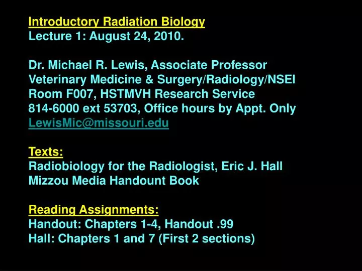
Introductory Radiation Biology Lecture 1: August 24, 2010. Dr. Michael R. Lewis, Associate Professor Veterinary Medicine & Surgery/Radiology/NSEI Room F007, HSTMVH Research Service 814-6000 ext 53703, Office hours by Appt. Only LewisMic@missouri.edu Texts:

E N D
Introductory Radiation Biology Lecture 1: August 24, 2010. Dr. Michael R. Lewis, Associate Professor Veterinary Medicine & Surgery/Radiology/NSEI Room F007, HSTMVH Research Service 814-6000 ext 53703, Office hours by Appt. Only LewisMic@missouri.edu Texts: Radiobiology for the Radiologist, Eric J. Hall Mizzou Media Handount Book Reading Assignments: Handout: Chapters 1-4, Handout .99 Hall: Chapters 1 and 7 (First 2 sections)
For Radiation Biology, our main interest is in the biological effects of ionizing radiation produced by artificial sources or radioactive decay processes. Many of you will be working with radiation sources and/or radioactive materials as a profession. It is essential that you understand not only the biological effects of radiation, but also the physical interactions of radiation with matter (which by in part give rise to the biological effects). Some radiations are more hazardous to one’s health than others, hence the reasoning for this understanding.
The Hazards from Ionizing Radiation Depend On the Following: • The type of radiation. • The mode of decay of the radionuclide. • The half-life of the radionuclide. • The biological handling of the radionuclide.
The History of Radiation Sciences/Radiobiology: • December 28 1895, Wilhelm Conrad Rontgen: Discovered a “new kind of ray” emitted from gas discharge tubes that blackened photographic plates in light-tight containers. He called them X-rays because of their unknown nature. • When his work was complete, X-rays were very well-characterized; he won the 1901 Nobel Prize in Physics for this discovery. • January 1896: Rontgen produced the first radiograph of a human hand. The first medical use of X-rays were to locate and remove a piece of knife blade from the backbone of a drunken sailor. • 1896, Henri Becquerel: Showed the existence of naturally-occuring radiaiton in uranyl sulfate; he won the 1903 Nobel Prize in Physics. • 1898, Pierre & Marie Curie: Concluded that uranium rays are an atomic phenomenon characteristic of the element and not related to it’s chemical or physical state. They named it “radioactivity” and shared the 1903 Nobel Prize in Physics with Becquerel. The Curies isolated Po & Rad from a ton of pitchblende and Marie won the 1911 Nobel Prize in Chemistry for discovery of these elements (Pierre had died in 1906 in a carriage accident).
Early Biological Effects of Radiation: • April 1896, Daniels: Daniels, a physician at Vanderbilt University, demonstrated the first biological effect of ionizing radiation (the epilation of scalp after X-irradiation of the skull). • 1897: Freund removed a hairy birthmark, considered to be the first therapeutic use of X-rays. • 1899, First Tumor Therapy: Stenbeck & Sjogren in Sweden removed a skin tumor from the tip of a patient’s nose with X-rays. • 1903, Senn: In Chicago, reduced the spleen sized of leukemic patient with X-rays.
Early Harmful Effects of Radiation: • Henri Becquerel: Becquerel used to carry a radium vial in his vest pocket for demonstrations. He noticed a reddening of the skin (erythema) in this area and the subsequent development of an ulcer. Pierre Curie reproduced this experiment in 1901 by deliberately producing a radium burn on his forearm, charting the appearance and healing of the radiation ulcer. Thus, the field of Radiobiology was born. • Harmful effects became obvious by the early 1900s, but went unattended for many years. • Latent Cancer: Both Marie & Irene Curie died of leukemia at an advanced age, probably the result of exposure to high levels of radioactivity. • Mutation: H. J. Muller (1927) demonstrated that ionizing radiation produced genetic mutations in the fruit fly Drosophila. L.J. Stadler (MU) showed the same effect in maize. Muller published ~2 months earlier than Stadler and was awarded the 1946 Nobel Prize in Physiology/Medicine.
Radiation Energetics: Two Classes of High Energy Radiation: • Particulate Radiation. • Electromagnetic Radiation. Properties of Particulate Radiation: • Mass: Particles are composed of matter. Particles have mass. • Kinetic Energy: • Classical Treatment: E = 1/2mv2 • Einstein: Mass-Energy Equivalence, E = mc2 (c = 3x10⁸ m/s) • Mass is a form of energy and mass and energy are interconvertible. Total mass + energy in any given process is constant.
3. Charged Particles: Charges on particles have associated electric and magnetic fields (wave-like properties). • Nomenclature: • Electron: e- = b- • Positron: e+ = b+ • Proton: 1H+ = p+ • Neutron: 1n = n • Deuteron: 2H+ = d+ • Alpha: 4He2+ = a2+
Properties of Electromagnetic Radiation (EMR): EMR is Often Called “Pure Energy” or “Light”. • Mass: EMR has no mass. • EMR is composed of oscillating electric & magnetic fields. • Charge: EMR has no charge. • Energy: EMR has energy. However, it is not considered to be classical kinetic energy. • EMR exhibits both wave-like and particle-like properties. • Wave Properties: EMR was first depicted in the form of a wave by Maxwell in 1864. • Maxwell stated we can describe light, or EMR by the “wave equation”, c = ln, where c = the speed of light, l = the wavelength, and n = the frequency of the oscillation (n is proportional to 1/l and l is proportional to 1/n. In other words, the shorter the wavelength, the higher the frequency.
EMR Undergoes the Following Wave-like Properties. • Optical Interference: • 2. Polarization: • Reflection: • Refraction: • Diffraction: • Wave Properties: All of this seemed to be a reasonable description until 1901. • 1901: Max Planck demonstrated that E was proportional to n, and that EMR could possess particle-like properties…, i.e., a “photon” or “quantum of light”. • Planck determined that E = hn, where h = Planck’s Constant.
Particle-like Properties of EMR Continued. EMR can be completely characterized by the following two mathematical equations. c = lnE = hn We need only to know one of the three variables to calculate the other two. E = hn = hc/l Important constants and values to consider: h = 6.6x10⁻²⁷ erg-s = 4.1x 10⁻¹⁵ eV-s 1 eV = 1.6x10⁻¹² erg 1 J = 107 erg (SI unit is the joule) c = 3x10⁸ m/s = 3x1010 cm/s Wavelength: 1 nm = 10-9m = 10-7cm 1 Angstrom (Å) = 0.1nm = 10-10m = 10-8cm
Concepts of Electromagnetic Radiation (EMR): • Units of Energy: We use the erg or the eV in this course; the traditional unit in Radiation Sciences is the eV. • Definition: One electron-volt (eV) is the kinetic energy gained by one electron when accelerated through a potential difference of one volt (V). • Biological Effects of EMR with energy of 1ev versus 1MeV: Must evaluate the chemical consequences of the action of EMR of various energies (See the EMR spectrum on page 19 of handout).
For l≥ 5-10cm up to miles in length: Radiowaves or microwaves with E < 10-3-10-10 eV. • For l≥ 700nm: Infrared with E = 0.01-1eV. • These type of radiations cause increases in the translational, rotational, and vibrational motion of molecules. The resulting increase in molecular energy is often converted to heat, as in a microwave oven or infrared heating lamp. • For l = 300-700nm: Visible spectrum withE = 1-2 eV.
In the visible portion of the spectrum, a single photon of light can now begin to initiate a chemical reaction by electronic excitation. Electrons in molecules can be excited to higher energy levels, where they become highly reactive. • However, this is the wavelength range to which we are continually exposed. Only a few, select, beneficial reactions are initiated above 380nm. For example… 11-cis-retinal is the reactive absorber in rhodopsin, the retinal rod cell pigment which has an absorption maximum of ~520 nm. Conversion to all trans-retinal form results in dissociation of retinal and a conformational change in opsin, which triggers a nerve impulse interpreted by the brain as sight.
Exercise: Calculate the energy of this photon (520 nm) in eV. • E = hn = hc/l = (4.1 x 10⁻¹⁵ ev-s)(3x10⁸m/s)/(520 x 10-9m) • E = 2.4 eV • Another beneficial reaction initiated by visible excitation is the absorption of 680nm light by chlorphyll. This induces electron transport processes that result in photosynthesis. • l = less than 200 to 380nm: UV light • 300-380nm: near UV • 200-300nm: UV • <200nm: vacuum UV • Between 250-310nm, 4-5eV, nucleic acids and proteins begin to absorb light.
Excitation: We are still in the energy range (UV) where molecular excitation occurs. Direct electronic excitation can damage nucleic acids & proteins, leading to mutations and cell death. Fortunately, ozone in the upper atmosphere acts as a protective absorber, screening out virtually all light below 290 nm. However, even UV with l = 290-350 nm has enough energy to cause substantial damage to cells; UV-A & UV-B in this range can cause sunburn and skin cancer. It is not until we get to the vacuum UV or X-ray range that ionization begins to occur.
Ionization Energy: The Ei of an atom or molecule is the amount of energy required to remove (to infinity) an electron from the atom or molecule isolated in free space and in its ground electronic state. Ionizing Radiation: Subatomic electronic particles or electromagnetic waves that are energetic enough to detach electrons from atoms or molecules. The ionization potential of most molecules is ~10eV. Ion Pair: A positively charged ion and a negatively charged ion produced by the addition of sufficient energy to a neutral atom or molecule to cause it to dissociate into oppositely charged fragments. Example: H2O H2O* H2O·+ + e- IP = 12.6 eV C6H6 C6H6* C6H6·+ + e- IP = 9.25 eV hn hn
X-rays and Gamma (g)-rays: • X-rays: Originate from orbital electron transitions or orbital electron rearrangement. Typically, X-rays between 10 and 11 keV are studied very little as they have limited penetrating ability. • g-rays: Originate from the unstable nucleus of an atom as it is undergoing decay. • We usually deal with X-rays or g-rays with E 11 keV. • As stated above, X-rays and g-rays have different points of origin. • We can describe X-rays and g-rays using the two equations for EMR. • Exercise: Calculate the energy of the characteristic X-ray of W. • KL transition of 0.2 Å = 2x10⁻⁹cm • E=hc/l = (4.1 x 10⁻¹⁵ eV-s)(3x1010cm/s)/2x10-9cm = 61500 eV (62keV) • THERFORE, SINCE THE IONIZATION POTENTIAL for MOST MOLECULES is ~10eV, YOU CAN SEE WHY g- and X-rays are TRULY IONIZING RADIATIONS!
THESE TYPES of RADIATIONS have SUFFICIENT ENERGY in ONE PHOTON to IONIZE MANY MOLECULES. Example: Co-60 has 2g photons (1.17 MeV and 1.33 MeV) of average photon energy of 1.25 MeV. How many molecules can a single g-photon of Co-60 ionize? 1.25x10⁶ eV/10eV/molecule = 125,000 molecules! However, the ionization process is not perfectly efficient. For example, 34eV of energy is deposited for each ion pair formed. If all of the energy were deposited, then: 1.25x10⁶ eV/34eV/molecule = 37,000 molecules would be ionized.
Thus, photons of X- or g-radiations represent large individual “packets” of energy. Each of these “packets” can ionize many molecules and initiate chemical events that lead to biological damage. A radiation dose that is lethal to a human being deposits the same amount of energy as the heat absorbed by drinking one sip of hot coffee. The critical difference is not the total energy involved, but the size of the energy “packets”.
Introductory Radiation Biology Lecture 2: August 26, 2010. Atomic Structure: The atom is composed of 3 subatomic particles • Proton (p+): mass = 1.007593 amu = 1.7 x 10⁻²⁴g • 2. Neutron (n): mass = 1.008982 amu = 1.7 x 10⁻²⁴g • Electron (e-): mass = 0.000549 amu = 9.1 x 10⁻²⁸g • mp+ = mn>> me- • Therefore, the atomic weights of all stable elements are known. • The electronic structure of the atom is described by the Bohr model. Electrons orbit the nucleus in distinct shells. Electrons can be excited into empty orbitals of higher energy. Transition back to a lower energy state results in the release of EMR (i.e., fluorescence, phosphoresence, X-rays).
Nuclide: Any nucles plus its orbital electrons. Defined as follows: AZX Where A is equal to the atomic mass (protons + neutrons), Z is equal to the atomic number (number of protons), and n = A-Z. Isotope: Is an atom of the same element, having the same number of protons, but differing numbers of neutrons. 116C, 126C, 136C, 146C Isomers: Atoms that have a constant atomic mass (A) and atomic number (Z), yet they contain a different energy arrangement of the nucleons. Example: 99m43Tc versus 99g43Tc
Binding Energy of Nucleons: Nucleons (p+, n) are held together in a very small volume of high positive charge density. Short-range nuclear forces overcome charge repulstion between protons. We can calculate the BE (binding energy) for nucleons). Remember, energy and mass are interconvertible (E = mc2). Dm = mtotal – mnucleus = BE Some of the mass of the individual p+ & n is converted to binding energy which holds the nucleus together. Therefore, the mass of the nucleus is less than the total mass of p+ + n.
Example (Binding Energy per Nucleon: Consider the 42He atom. Dm = mtotal – mnucleus = BE Mass of Nucleus: Measured atomic mass = 4.003873 amu Mass of 2 electrons = 0.001098 amu Mass of Nucleus = 4.002775 amu Mass total: 2 x mp+ = 2 x 1.007593 = 2.015186 amu 2 x mn = 2 x 1.008982 = 2.017964 amu Mass total = 4.033150 amu Dm = mtotal – mnucleus = BE Dm = 4.033150 – 4.002775 Dm = 0.030375 amu Mass Energy: 1amu = 931 MeV So, Binding Energy (BE) = 0.030375 amu (931MeV/amu) = 28.28 MeV Binding Energy per Nucleon = 28.28 MeV/4 nucleons = 7.07 MeV/nucleon
Various models of nuclear structure exist. One thing is very certain; there are quantized energy levels which nucleons occupy in the nucleus. We can consider this similar in concept to the arrangement of electrons in atomic orbitals. Properties of Stable Nuclei:Systematic investigations of nuclei indicate some general rules regarding nuclear stability. • If there is more than one proton (p+) in the nucleus, then there must be some neutrons. • 2. There are no stable nuclides with Z > 83. 20983Bi is the heaviest element with stability. This is the maximum number of nucleons (p + n) that can be packed in the nucleus and have stability. • Even numbers of nucleons (p or n) are preferred. Only 4 stable odd-odd nuclides exist: 21H, 63Li, 105B, 147N. • Stable nuclides appear to cluster around a line of stability. For example, for each Z, there appears to be only certain numbers of n that will result in stability.
Line of Beta Stability: Thus, very often, unstable nuclei are either proton rich or neutron rich. Also, odd-odd combinations can prompt instability
5. “Magic Numbers” which result in rather exceptional stability do exist. • Either N or Z = 2, 8, 20, 28, 50, 82, 126 • These are closed shells, analogous to those seen in electronic structure 2n2 e-.
Modes of Radioactive Decay: • Alpha Decay: Fast moving monoenergetic helium nuclei. This decay only occurs in elements of very high molecular weight. Decrease of 4 in atomic mass (A) and 2 in atomic number (Z). • Negatron (b-): Negatively charged electrons released from the nuclei of neutron rich radionuclides (n p + b- + antineutrino).Increase of +1 in atomic number. • Positron (b+): Positively charged electrons are released from the nuclei of proton rich radionuclides. (p n + b+ + neutrino).No change in mass number, but decrease of 1 in atomic number. • Gamma Ray Decay: The nucleus rids itself of excess energy by the emission of photons (all having the same discrete energy). When gamma emission takes place between two states of measurable lifetime, gamma decay is called isomeric transition. In gamma decay, the number of nucleons does not change! • Electron Capture (EC): Occurs when a K orbital electron interacts with the nucleus, combining with a proton to form a neutron and a neutrino (e- + p n + neutrino). Loss of 1 proton gain of 1 neutron per nucleus. • Internal Conversion: In most modes of decay where gamma rays are normally emitted, the excited nucleus may interact with an inner orbital electron, with all of the excitation energy being transferred to that electron. The electron will be ejected from the atom. Conversion electrons are monoenergetic.
Alpha Decay: Fast moving monoenergetic helium nuclei. This decay only occurs in elements of very high molecular weight. Decrease of 4 in atomic mass (A) and 2 in atomic number (Z). An alpha particle is a 42He atom: The radioactive decay mechanism is as follows: AZX A-4Z-2Y + 42He + g + Q Q value: The energy release by the decay process. It is equal to the mass of the parent nucleus minus the mass of the decay products. If it is a positive number, decay can occur. Radioactive Decay: Spontaneous release of energy in the form of subatomic particles or EMR in an attempt to reach a more stable state.
Example: Radium-226 decay. 22688Ra, t1/2 = 1620y (a2 = 4.60MeV, 6.5%) (a1 = 4.78MeV, 93.5%) (g = 0.186MeV) 22286Rn, t1/2 = 3.8d Arrows go to the left. A decreases by 4 and Z decreases by 2.
Example of Q: Consider Pathway for a 1 (93.5%). Q =Dm(931 MeV/amu) = (mp - (mD + ma)(931MeV/amu) Mass of Parent = 226.0254 amu Mass of Daughter = 222.0175 amu Mass of Alpha = 4.0027 amu Change in mass: Dm = 226.0254 – (222.0175 + 4.0027) Dm = 0.0052 amu Mass Energy: 1amu = 931 MeV Q = 0.0052 amu (931MeV/amu) = 4.87 MeV
Example of Q: Consider Pathway for a 2 (6.5%). Q =Ea + Eg+ Recoil E Recoil E = (ma/mrecoil)Ea1 = (4/222)4.78MeV = 0.086MeV Q = 4.60MeV + 0.186MeV + 0.086 MeV = 4.87MeV Summary: Ra-226 decay. Ra-226 loses a 42He atom during decay. Energy is also released in the form of KE of the 42He atom. The emission is monoenergetic, indicating the existence of discrete energy levels in the nucleus. Q cannot vary from parent to daughter. The total energy loss from parent to daughter is ALWAYS the same! a2 decay allows the nucleus to go to an intermediate energy level (i.e., 0.186MeV above ground state). Then the nucleus eliminates the remainder of the energy (sometime later, ~1ns) as EMR (g photon).
The gamma photon actually comes from the daughter nucleus (Rn). So, from this example, you can see that there is more than a single pathway by which E is lost. Sometimes, radioactive decay can leave the daughter nucleus in an “excited state”. The release of E from this “excited state” occurs via g-photon emission.
2. Negatron (b-): Negatively charged electrons released from the nuclei of neutron rich radionuclides (n p + b- + antineutrino).Increase of +1 in atomic number. A negatron is a 0-1e- (b-): The radioactive decay mechanism is as follows: AZX AZ+1Y + 0-1e + n + g + Q
Example: Phosphorus-32 decay. 3215P, t1/2 = 14d (b = 1.72MeV, 100%) 3216S, stable Arrow goes to the right. Note a net increase in Z of 1 (neutron to a proton. Q = 1.72 MeV
Example of Q: Consider Pathway for b 1 (100%). Q =Dm(931 MeV/amu) = (mp - (mD)(931MeV/amu) Mass of Parent = 31.98412 amu Mass of Daughter = 31.98227amu Mass of Electron = Already accounted for because Z increases by 1 Change in mass: Dm = 31.98412 – (31.98227) Dm = 0.00185 amu Mass Energy: 1amu = 931 MeV Q = 0.00185 amu (931MeV/amu) = 1.72 MeV
With an energy of 1.72 MeV, we would expect the b- to be monoenergetic. However, this is not the case. Rather, a continuum of b- energy is observed up to the maximum: Se the graph below. Typically, Eb-(average) ~ 1/3 Emax.
To avoid the necessity of abandoning conservation laws (E & spin), we postulate another particle, the antineutrino (n), which has zero charge, zero mass, and a spin of 1/2. The antineutrino carries away the excess decay energy not imparted to the b-. Q = Eb- + En + Erecoil Summary of Beta Decay: Results in transforming a neutron to a proton in the nucleus. An antineutrino is emitted in conjugation with the b- particle. Even though the Etotal for the decay is always the same, Eb- is a contnuum with Eb-(average) ~ 1/3 Emax. Beta decay occurs in neutron rich radionuclides. Gamma emission can accompany b- decay.
6027Co, t1/2 = 5y (b = 0.32MeV, 100%) g1 = 1.17MeV g1 = 1.33MeV 6028Ni, stable