Download
1 / 7
70 likes | 83 Views
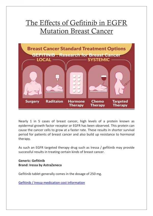
The precise causes of psoriasis, a relatively common, chronic, inflammatory and hyperproliferative skin disease, are<br>still not known, thus making it very difficult for treatment. We previously found an in vitro anti-psoriatic activity in<br>ethanolic extract of Annona squamosa L. leaves. Epidermal growth factor receptor (EGFR) and its ligand,<br>transforming growth factor (TGF)-α, have been demonstrated to be strikingly upregulated in active psoriatic<br>plaques, thus implicating their functional roles in psoriatic hyperplasia. The objective of this study was to<br>investigate the molecular effect of ethanolic extract derived from Annona squamosa L. leaves on the EGFR<br>expression using HaCaT keratinocyte cell line as a model. Based on RT-PCR, concentrations at 3.15 and 1.575<br>µg/mL (IC50= 6.3 µg/mL) significantly reduced the EGFR mRNA expression (P<0.05) as compared to control<br>(HaCaT pretreated with IFN-γ and TNF-α, 10 ng/mL for each) although all three concentrations tested showed a<br>similar tendency of inhibitory effect. Using confocal immunofluorescence microscopy as a qualitative measurement,<br>we also found the inhibitory effect of Annona squamosa L. leaf extract on the expression of EGFR protein. Western<br>blot analysis confirmed that all concentrations of the extract tested markedly inhibited the expression of EGFR<br>(P<0.05). Taken together, this suggest that the ethanolic extract of Annona squamosa leaves could exert its<br>biological effect by suppressing the expression of EGFR biomarker, thus highlighting the potential therapeutic<br>development of this extract for future applications.

E N D
Available online www.jocpr.com Journal of Chemical and Pharmaceutical Research, 2014, 6(4):791-797 ISSN : 0975-7384 CODEN(USA) : JCPRC5 Research Article Effects of ethanolic extract of Annona squamosa L. leaves on the expression of EGFR Chalinee Ronpirin1, Thitiporn Charueksereesakul2, Visa Thongrakard2 and Tewin Tencomnao3* 1Preclinical Science, Faculty of Medicine, Thammasat University, Pathumthani, Thailand 2Graduate Program in Clinical Biochemistry and Molecular Medicine, Department of Clinical Chemistry, Faculty of Allied Health Sciences, Chulalongkorn University, Bangkok, Thailand 3Center of Excellence in Omics-Nano Medical Technology Development Project, Department of Clinical Chemistry, Faculty of Allied Health Sciences, Chulalongkorn University, Bangkok, Thailand ____________________________________________________________________________________________ ABSTRACT The precise causes of psoriasis, a relatively common, chronic, inflammatory and hyperproliferative skin disease, are still not known, thus making it very difficult for treatment. We previously found an in vitro anti-psoriatic activity in ethanolic extract of Annona squamosa L. leaves. Epidermal growth factor receptor (EGFR) and its ligand, transforming growth factor (TGF)-α, have been demonstrated to be strikingly upregulated in active psoriatic plaques, thus implicating their functional roles in psoriatic hyperplasia. The objective of this study was to investigate the molecular effect of ethanolic extract derived from Annona squamosa L. leaves on the EGFR expression using HaCaT keratinocyte cell line as a model. Based on RT-PCR, concentrations at 3.15 and 1.575 µg/mL (IC50= 6.3 µg/mL) significantly reduced the EGFR mRNA expression (P<0.05) as compared to control (HaCaT pretreated with IFN-γ and TNF-α, 10 ng/mL for each) although all three concentrations tested showed a similar tendency of inhibitory effect. Using confocal immunofluorescence microscopy as a qualitative measurement, we also found the inhibitory effect of Annona squamosa L. leaf extract on the expression of EGFR protein. Western blot analysis confirmed that all concentrations of the extract tested markedly inhibited the expression of EGFR (P<0.05). Taken together, this suggest that the ethanolic extract of Annona squamosa leaves could exert its biological effect by suppressing the expression of EGFR biomarker, thus highlighting the potential therapeutic development of this extract for future applications. Keywords: Psoriasis, Annona squamosa, EGFR, HaCaT, Mechanistic effect of herbal extract ____________________________________________________________________________________________ INTRODUCTION Psoriasis is a common chronic skin disease, characterized by abnormal keratinocyte proliferation, affecting approximately 1-3% of population worldwide [1]. Psoriatic patients have upregulated expression levels of various markers including angiogenic peptides, growth factors, receptors and cytokines, thus mediating certain signaling cascades to result in hyperproliferation and abnormal differentiation of keratinocytes [2]. The pathogenesis of psoriasis remains to be elucidated; it is however recognized as an autoimmune-associated disease with unidentified infectious agents or antigens [3]. Patients with psoriasis depend on a long-term treatment, which significantly affects their quality of life. Specifically, in addition to certain side effects of the treatment, they may face not only an increase in expense, but also a decrease in employment and income. The pathogenesis of psoriasis is not exactly known, thus making it very difficult for effective therapy. Recently, there was a study revealing a prolonged positive effect of erlotinib, the tyrosine kinase of epidermal growth factor receptor (EGFR) commonly used in lung cancer treatment, on psoriasis in patients [4]. Those cases were actually suffered from lung cancer and concomitant psoriasis. The unexpected outcome of treatment with 791
Chalinee Ronpirin et al. ______________________________________________________________________________ J. Chem. Pharm. Res., 2014, 6(4):791-797 erlotinib was subsequently of a great interest in the field of dermatology. Indeed, EGFR is essential in normal skin development and function. Both transforming growth factor-α (TGF-α) [5, 6] and its receptor, EGFR [7, 8] have been found to be upregulated in active psoriatic plaques, thus implicating their functional roles in psoriatic hyperplasia. Consistently, transgenic gene expression of the human amphiregulin, a member of the EGF system, was demonstrated to induce a psoriasis-like phenotype [9]. Therefore, EGFR has been proven to be a molecular target for therapy of psoriasis. Previously, we demonstrated that dithranol, a well-established agent known as anti-psoriatic drug, suppressed the EGFR mRNA expression of human HaCaT keratinocyte cell line, an in vitro model for psoriasis, in a dose- dependent fashion [10]. Our finding was in line with the early finding that another known anti-psoriatic agent, vitamin D3 derivative, inhibited the expression of EGFR in human epidermal keratinocytes [11]. Current treatments for psoriasis not only lack effective outcomes, but also result in side effects. It is our interest to discover alternative medicine to satisfy the psoriatic patients. Herbal medicine should be further explored as an optional treatment for psoriasis. Recently, indirubin, known as a natural plant compound, was demonstrated to downregulate the EGFR expression [12]. Flavonoid quercetin from the rhizome of Smilax china Linn. was shown to possess anti-psoriatic activity [13]. According to our previous investigation, we found three ethanolic herbal extracts from eleven medicinal plant species possessing in vitro anti-psoriatic activity, Alpinia galanga L. (rhizome), Curcum longa L. (rhizome) and Annona squamosa L. (leaf) [14]. It was lately demonstrated that the ethanolic extract of Annona squamosa L. leaves was rich in quercetin [15]. Only quercetin, not rutin, was displayed to exhibit anti-psoriatic activity although both chemicals are structurally related [16]. In the current study, we examined whether ethanolic extract of Annona squamosa L. leaves exhibited its anti-psoriatic effect via EGFR signaling. EXPERIMENTAL SECTION Plant materials Annona squamosa was collected from the Princess Maha Chakri Sirindhorn Herbal Garden (Rayong Province, Thailand). It was authenticated by Professor Dr. Thaweesakdi Boonkerd (Department of Botany, Faculty of Science, Chulalongkorn University, Thailand). The voucher specimens [013399 (BCU)] was deposited at the Professor Kasin Suvatabhandhu Herbarium, Department of Botany, Faculty of Science, Chulalongkorn University, Thailand. Preparation of Annona squamosa L. leaf extract Annona squamosa L. leaves were extracted by maceration at room temperature with ethanol (Merck, Hohenbrunn, Germany) using a 1:5 (w/v) ratio in a shaking incubator at 120 rpm for 48 h. The medicinal plant extract was filtered, and the residues were subsequently extracted twice more. After the two filtrates were pooled, the crude extract was concentrated using the MiVac Quattro concentrator at 45 °C. Finally, the resulting crude extract was dissolved in dimethyl sulphoxide (DMSO, Merck) as stock solutions (100 mg/mL), stored at −20 °C and protected from light. Prior to incubation with HaCaT cells, the crude extract was filtered through a 0.2 µm pore size filter (Corning Inc., Corning, NY, USA). Cell culture and treatment of HaCaT cells HaCaT cells, purchased from Cell Lines Service (CLS, Heidelberg, Germany), were seeded 3x10⁶ cells/mL in 15x60 mm of Petri dish and grown in Dulbecco’s Modified Eagle Medium/high glucose supplemented with 10% fetal bovine serum, 100 U/mL penicillin and 100 µg/mL streptomycin (Hyclone, Logan, UT, USA) at 37 ºC in 5% CO2. The cells were pretreated with pro-inflammatory cytokines, IFN-γ (10 ng/mL) and TNF-α (10 ng/mL) (Peprotech, Rocky Hill, NJ, USA) for 24 h. Subsequently, HaCaT cells were treated with various concentrations of ethanolic extract: 6.3, 3.15, 1.575 µg/mL (IC50= 6.3) of Annona squamosa L. leaf extract for the period of 48 h at 37 ºC in 5% CO2. Controls were performed with DMSO and medium alone. Each sample concentration was tested in triplicates. Semi-quantitative reverse transcriptase-polymerase chain reaction (RT-PCR) Prior to total RNA extraction from HaCaT cells, cells were washed with PBS. Isolation of total RNA was performed using Trizol reagent following the manufacturer’s instructions (Invitrogen, Carlsbad, CA, USA). Before performing the RT reaction, total RNA was adjusted to the final concentration of 1 μg/uL, and RNA was treated with 10−5 U deoxyribonuclease I (DNase I) (Invitrogen) for 30 min at 37 °C. DNase I-treated RNA was reverse transcribed by ImProm-IITM Reverse Transcription System (Promega, Madison, WI, USA) with oligo(dT)17 primer. For amplification of the cDNA, each desired DNA fragment was amplified for 35–40 cycles using each gene- specific primer pair for EGFR (forward primer, 5’ GCT ACG ATT GGC TGA AGT AC 3’ and reverse primer, 5’ ATT GGG TGT AGA GAG ACT GGA 3’) [17] and β-actin (forward primer, 5’ ACG GGT CAC CCA CAC TGT GC 3’ and reverse primer, 5’ CTA GAA GCA TTT GCG GTG GAC GAT 3’) [18] with expected product sizes of 518 bp and 656 bp for EGFR and β-actin, respectively. The products of RT-PCR were resolved by 2% agarose gel 792
Chalinee Ronpirin et al. ______________________________________________________________________________ J. Chem. Pharm. Res., 2014, 6(4):791-797 electrophoresis and visualized by ethidium bromide staining. All band densities were analyzed by the GeneTools program in a G:Box(Syngene, Cambridge, UK). Confocal immunofluorescence microscopy The HaCaT cells were seeded 1x10⁵ cells/mL on coverslip in a 6-well plate at 37 ºC in 5% CO2. The cells were pretreated with 10 ng/mL IFN-γ and 10 ng/mL TNF-α for 24 h. Subsequently, the cells were treated with various concentrations of Annona squamosa L. leaf extract for 48 h at 37 ºC in 5% CO2. Cells were washed with cold PBS three times and fixed with absolute methanol at -20 °C for 5 min. Fixed cells were washed with PBS and then permeabilized by adding 0.5% Triton X-100 with 0.05% Tween20 in PBS for 1 h at room temperature. Cells were exposed to primary antibody (anti-EGFR, Cell Signaling Technology, Danvers, MA, USA), and detection using anti-rabbit Alexa 555 (Cell Signaling Technology) was performed. After staining of EGFR-positive cells, cells were washed for 5 min in PBS three times and stained the nucleus with Hoechst 33258 for 15 min at room temperature. After washing for 5 min in PBS three times, cells were observed using a confocal laser scanning microscope (LSM 700, Carl Zeiss, Germany). Protein extraction and Western blotting analysis Total protein isolation from the HaCaT cells was carried out using Nonidet P40 lysis buffer. Protein concentrations were determined using the Bradford method [19]. The protein samples were frozen at -80 °C for later analyses. For Western blotting, thirty µg of protein extracts were separated by 10% sodium dodecyl sulfate (SDS)-polyacrylamide gel electrophoresis and transferred onto a polyvinyldine fluoride (PVDF) membrane. The membrane was then blocked with 5% non-fat dry milk in Tris-buffered saline Tween20 and incubated with either polyclonal rabbit anti- EGFR (1: 10,000, Cell Signaling Technology) or polyclonal rabbit anti-GAPDH (1: 10,000, Santa Cruz Biotechnology, Santa Cruz, CA, USA) for 1 h. After washing, the membrane was incubated with anti-rabbit IgG, horseradish peroxidase-linked secondary antibody, diluted at an appropriate dilution in 5% BSA, for 45 min at room temperature. The expected protein bands, EGFR (175 kDa) and GAPDH (37 kDa), were visualized using an enhanced chemiluminescence system with SuperSignal West Femto Maximum Sensitivity Substrate (Thermo scientific, Rockford, IL, USA). The results were normalized to GAPDH values. Statistical analysis All values are expressed as means ± SEM. The data were analyzed by the Student t-test. P < 0.05 was considered to be significant. Each experiment consisted of at least three replicates per condition. RESULTS AND DISCUSSION The ethanolic extract of Annona squamosa L. leaves was previously demonstrated by our group to contain anti- psoriatic activity with IC50 of 6.3 µg/mL using the HaCaT cell line as an in vitro psoriatic model for [14]. Currently, this particular model has been accepted as a suitable model [10]. Specifically, to make this HaCaT cell line a psoriatic model, the cells have been recommended to be pretreated with 10 ng/mL IFN-γ and 10 ng/mL TNF-α for 24 h. Therefore, this pretreated condition was used as a control in this study. Certain ethanolic extracts of Thai plant species have been elucidated to exhibit anti-psoriatic effect via a few possible mechanisms such as increase in apoptosis via caspase 9 [20] and decrease in inflammation via NF-κB signaling pathway [21]. In this in vitro study, we aimed at elucidating the molecular mechanism to demonstrate whether the ethanolic extract of Annona squamosa L. leaves could exert its anti-psoriatic effect by suppressing the expression of EGFR. The EGFR plays a vital role in development, differentiation, and motility of normal as well as cells in hyperproliferative states. For predictive disease diagnostics and therapeutic targeting of EGFR, it is critical to know how the EGFR signaling pathway is regulated. We started out by determining the effect of the ethanolic extract of Annona squamosa L. leaves on the expression of EGFR mRNA. As shown in Figure 1, the EGFR mRNA expression was significantly reduced by RT-PCR (P < 0.05) when HaCaT cells were treated with 1.575 µg/mL (0.25IC50) and 3.15 µg/mL (0.5IC50) of Annona squamosa L. leaf extract. Nevertheless, we did not observe the significance of inhibitory effect of this extract at the highest concentration tested, 6.3 µg/mL (IC50). In fact, there was a tendency toward reducing the expression of EGFR mRNA by this highest concentration of the extract as compared to the control. 793
Chalinee Ronpirin et al. ______________________________________________________________________________ J. Chem. Pharm. Res., 2014, 6(4):791-797 A A A B B B Figure 1. mRNA Expression of EGFR and β-actin as detected by RT-PCR in HaCaT cells when treating cells with 6.3, 3.15 or 1.575 µg/mL (IC50= 6.3) of Annona squamosa L. extract for 48 h. A. The expression levels of EGFR and β-actin were displayed as a result of RT-PCR. Lanes 1: 100 bp ladder, 2: HaCaT cells with IFN-γ and TNF-α (control), 3: HaCaT cells with IFN-γ, TNF-α and 0.0067% DMSO, 4: HaCaT cells with IFN-γ, TNF-α and 1.575 µg/mL (0.25IC50) of Annona squamosa L., 5: HaCaT cells with IFN-γ, TNF-α and 3.15 µg/mL (0.5IC50) of Annona squamosa L., 6: HaCaT cells with IFN-γ, TNF-α and 6.3 µg/mL (IC50) of Annona squamosa L., 7: Negative control of RT, 8: Negative control of PCR. B. After EGFR mRNA is normalized against that of β-actin data represent the foldexpression over that of control. A symbol, *, indicating a statistically significant difference (P < 0.05). Although it was difficult to explain as we could not find a dose-dependent effect with regard to our mRNA experiment, we decided to subsequently examine the effect of Annona squamosa L. leaf extract on the expression of EGFR protein using confocal immunofluorescence study. Although a qualitative analysis was carried out, all three concentrations of the extract clearly suppressed the expression of EGFR protein as compared to the control (Figure 2). In addition to the expression level, transition of staining pattern was detected as untreated cells showed cytoplasmic staining of EGFR, but extract-treated cells showed intercellular staining. To confirm the expression level of EGFR protein, Western blotting was performed. As displayed in Figure 3, the expression of EGFR protein was markedly diminished by all three concentrations of the Annona squamosa L. leaf extract (P < 0.05), thus indicating the inhibitory effect of this herbal extract on the EGFR expression of our in vitro model, HaCaT cell line. The tyrosine kinase of EGFR protein is involved in many cancer types, and it is believed to play a vital role in the pathogenesis of psoriasis. This has made EGFR more interesting as a therapeutic target for cancer and psoriasis. Although quercetin might be the prime compound found in Annona squamosa L. leaves playing this suppressive effect on EGFR expression, we could not rule out the importance of other chemical constituents in the Annona squamosa L. leaf extract. Interestingly, in silico screening of EGFR in the tyrosine kinase domain through a medicinal plant compound database (29,960 compounds from 79 plant species) showed that one of eight potential hits was referred to as compound no. 113866-89-0 derived from Annona squamosa L. [22]. This particular compound was classified into phenylated flavone. This highlights the potential applications of Annona squamosa L. leaf extract and should be further explored for drug discovery. 794
Chalinee Ronpirin et al. ______________________________________________________________________________ J. Chem. Pharm. Res., 2014, 6(4):791-797 Figure 2. Expression of EGFR protein as detected by immunocytochemistry and confocal microscopy in HaCaT cells when treating cells with 6.3, 3.15 or 1.575 µg/mL (IC50= 6.3)of Annona squamosa L. extract for 48 h. Untreat: HaCaT cells treated with IFN-γ and TNF-α, 0.25IC50: HaCaT cells with IFN-γ, TNF-α and 1.575 µg/mL (0.25IC50) of Annona squamosa L., 0.5IC50: HaCaT cells with IFN-γ, TNF-α and 3.15 µg/mL (0.5IC50) of Annona squamosa L., IC50: HaCaT cells with IFN-γ, TNF-α and 6.3 µg/mL (IC50) of Annona squamosa L. 795
Chalinee Ronpirin et al. ______________________________________________________________________________ J. Chem. Pharm. Res., 2014, 6(4):791-797 A B EGFR 175 kDa Figure 3. Expression of EGFR and GAPDH proteins as detected by Western blotting analysis in HaCaT cells when treating cells with 6.3, 3.15 or 1.575 µg/mL (IC50= 6.3) of Annona squamosa L. extract for 48 h. A. The expression levels of EGFR and GAPDH were displayed as a result of Western blotting analysis. Lanes 1: HaCaT cells with medium alone (unstimulated condition), 2: HaCaT cells with IFN-γ and TNF-α (control or stimulated condition), 3: HaCaT cells with IFN-γ, TNF-α and 0.0067% DMSO, 4: HaCaT cells with IFN-γ, TNF-α and 1.575 µg/mL (0.25IC50) of Annona squamosa L., 5: HaCaT cells with IFN-γ, TNF-α and 3.15 µg/mL (0. 5IC50) of Annona squamosa L., 6: HaCaT cells with IFN-γ, TNF-α and 6.3 µg/mL (IC50) of Annona squamosa L. B. After EGFR protein is normalized against that of GAPDH data represent the foldexpression over that of control. A symbol, *, indicating a statistically significant difference (P < 0.05). CONCLUSION In conclusion, the inhibitory effect of ethanolic extract derived from Annona squamosa L. leaves on the EGFR expression was found using HaCaT keratinocyte cell line as a model. RT-PCR, confocal immunofluorescence microscopy and Western blotting consistently showed the reduced expression of EGFR as compared to control in response to the treatment with varying concentration of the Annona squamosa L. leaf extract.This effect was actually in accordance with erlotinib, the tyrosine kinase of EGFR as addressed in the previous report. Taken together, this might suggest that the ethanolic extract of Annona squamosa leaves could exert its inhibitory effect by targeting the expression of EGFR biomarker, thus highlighting the potential therapeutic development of this extract for future applications. Acknowledgments This research was supported by the grants from Thammasat University (2011) and the Royal Thai Government (2012) for C.R. and T.T., respectively. T.C. received tuition fee scholarship and teaching assistant fellowship from the Faculty of Allied Health Sciences, Chulalongkorn University. We gratefully acknowledge the generous supplies of Thai medicinal plants from the Princess Maha Chakri Sirindhorn Herbal Garden. 796
Chalinee Ronpirin et al. ______________________________________________________________________________ J. Chem. Pharm. Res., 2014, 6(4):791-797 REFERENCES [1] E Christophers. Clin. Exp. Dermatol., 2001, 26 (4), 314–20. [2] CO Mendonca; AD Burden. Pharmacol. Ther., 2003, 99(2), 133–47. [3] A Davidson; B Diamond. N. Engl. J. Med., 2001, 345(5), 340–50. [4] TR Overbeck; F Griesinger. Dermatology, 2012, 225(2), 179–82. [5] AB Gottlieb; CK Chang; DN Posnett; B Fanelli; JP Tam. J. Exp. Med., 1988, 167(2), 670–5. [6] JT Elder; GJ Fisher; PB Lindquist; GLBennett; MR Pittelkow; RJ Jr Coffey; L Ellingsworth; R Derynck ; JJ Voorhees. Science, 1989, 243(4892), 811–4. [7] LB Nanney; CM Stoscheck; M Magid; LE King Jr., J. Invest. Dermatol.1986, 86(3), 260–5. [8] YY Choi; KS Lee; SS Lee; ES Chang; JY Song, Korean Journal of Dermatology, 1992, 30(4), 431–8. [9] PW Cook; M Piepkorn; CH Clegg; GD Plowman; JM DeMay; JR Brown; MR Pittelkow. J. Clin. Invest., 1997, 100(9), 2286-94. [10] T Tencomnao; C Ronpirin; A Prasansuklab; Y Poovorawan, Afr. J. Biotechnol., 2009, 8(14), 3141–6. [11] AM Boisseau-Garsaud; P Donatien; C Margerin; A Taieb, Arch. Dermatol. Res., 1996, 288(8), 453–7. [12] WL Hsieh; YK Lin; CN Tsai; TM Wang; TY Chen; JH Pang, J. Dermatol. Sci., 2012, 67(2), 140–6. [13] A Vijayalakshmi; V Ravichandiran; V Malarkodi; S Nirmala; S Jayakumari,Asian Pac. J. Trop. Biomed., 2012, 2(4), 269-75. [14] V Thongrakard. Master’s degree thesis. Chulalongkorn University, Bangkok, Thailand,2009. [15] V Varadharajan; UK Janarthanan; V Krishnamurthy. WJPR, 2012, 1(4), 1143–1164. [16] SC Shen; WR Lee; LY Yang; HH Tsai; LL Yang; YC Chen, Exp. Dermatol., 2012, 21(5), 370–5. [17] N Kubota;Y Kiuchi; M Nemoto; H Oyamada; M Ohno;H Funahashi;S Shioda; K Oguchi, Eur. J. Pharmacol., 2001, 417(1-2), 69-76. [18] S Fisker; B Hansen; J Fuglsang; K Kristensen; P Ovesen; H Ørskov; JO Jørgensen Eur. J. Endocrinol., 2004, 150(6), 773-7. [19] MM Bradford, Anal. Biochem.1976, 7(72), 248-54. [20] T Charueksereesakul; V Thongrakard; T Tencomnao; J. Chem. Pharm. Res., 2011, 3(4), 196–203. [21] C Saelee; V Thongrakard; T Tencomnao; Molecules, 2011, 16(5), 3908–32. [22] O Sawatdichaikul; S Hannongbua; C Sangma; P Wolschann; K Choowongkomon; J. Mol. Model., 2012,18(3), 1241–54. 797