Download
1 / 30
370 likes | 961 Views
Wounds: Care and Treatment. William R. Dougherty, MD FACS Associate Professor of Surgery. Goals. Identify elements of wound healing Define differences between acute and chronic wounds Treatment options of acute wounds Treatment options of chronic wounds. Healing and Regeneration.

E N D
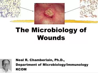
Wounds: Care and Treatment William R. Dougherty, MD FACS Associate Professor of Surgery
Goals • Identify elements of wound healing • Define differences between acute and chronic wounds • Treatment options of acute wounds • Treatment options of chronic wounds
Healing and Regeneration • Starfish and salamanders heal by regeneration (whole limbs) • Mammals heal by a combination of scarring and regeneration.
Surgeons Invocation Give us the ability to operate on the things that cannot be healed To heal the things that can be healed, And the wisdom to know the difference.
Acute Wounds • Acute wounds normally heal in a very orderly and efficient manner. • Healing is characterized on a four distinct but overlapping faces. • Hemostasis, Inflammation, Proliferation, and Remodeling • Specific biologic markers characterize and orchestrate the phases healing of the acute wounds.
Cytokines • Originally identified cell growth factors, now recognized to have diverse effects. • Endocrine, autocrine, paracrine and intracrine. • Cell migration, matrix production, enzyme expression and cell differentiation
Hemostatic Phase • Platelets come into contact with collagen and extracellular matrix. • Contact triggers the release of clotting factors as well as essential growth factors and cytokines. • PDGF platelet derived growth factor • TGFβ transforming growth factor beta
Inflammatory Phase • Neutrophils enter the wound site first to remove foreign materials, bacteria and image tissue. • Macrophages continue the process of phagocytosis and release more PDGF and TGFβ • Rubor, Tumor, Calor and Dolor
Proliferative Phase • In response to PDGF and TGFβ fibroblasts migrate in to deposit collagen and extracellular matrix. • College is the most abundant protein in the animal kingdom, accounting for 30% of the total protein in the human body. • Epithelial migration and new vessel formation are prominent features.
Proliferative Phase • Hydroxyproline is key in maintaining tertiary college and structure. • Vitamin C deficiency and hypoxia both reduce hydroxyproline • Lysyl oxidase (inhibited by steroids) is responsible for cross-linking collagen from
Collagen • Type I - Located in all connective tissue except hyaline cartilage and basement membranes • Type II - Located in hyaline cartilage • Type III - Located in distensible connective tissue (blood vessels) • Type IV - Located in basement membranes • Type V - Located in all tissues • Type VI - Located in all tissues • Type VII - Located in the dermal-epidermal junction • Type VIII - Located in the Descemet membrane • Type IX - Located in hyaline cartilage • Type X - Located in hypertrophic cartilage and hyaline cartilage
Remodeling Phase • The new collagen matrix is metabolized, cross-link and organized. • Native collagen approaches the strength of steel on a weight per weight basis • Defects in remodeling main result in low tensile strength or hypertrophic scaring • The healed wound reaches 80% of the surrounding normal tissue strength in six to eight weeks
Normal Wound Healing Scar maturation Collagen fibril cross-linking REMODELING Endothelial cells Epithelial cells Collagen Fibroblasts PROLIFERATION Lymphocytes Macrophages Neutrophils INFLAMMATION Proteoglycans Fibrin Platelets HEMOSTSIS Time from injury Wound healing phases: Overview
Factors that influence wound healing • Nutrition • Bacterial contamination • Necrotic tissue / debris • Immune / Bone marrow competence • Edema • Underlying medical conditions • Drug therapy • Radiation therapy
What’s new: Stem Cells • Bone marrow derived stem cells • Circulating and fixed • Responders to the cell signaling • Regenerative components
Acute Wounds: Etiology • Trauma • Burns • Vascular • Immune • Surgical • Pressure • Infection
Chronic Wounds • Unique biologic markers characterize pathologic healing responses that resulting fibrosis and chronic nonhealing wounds. • The efficient and orderly processes lost and the wounds are locked in to the state of chronic inflammation and fibrosis. • This is associated with abundant neutrophil infiltration, reactive oxygen species and district in enzymes.
Fibrosis • In most conditions of fibrosis are characterized by an increased density of mast cells. Mast cells contain specialized enzymes capable of processing procollagen and it has been suggested that abnormal peptides are produced that can actually stimulate collagen synthesis thus producing fibrosis.
Keloid versus Hypertrophic Scar • There is one very significant biological marker that distinguishes keloids from hypertrophic scars and that is the absence of myofibroblasts in keloids and an abundance of these contractile cells in hypertrophic scars.
Common Factors • Loss of epithelial integrity • Devascularized tissue • Contamination • Stimulation of cytokine cascade • Stimulation of stem cell migration • Coagulation, Inflammation, Proliferation and Remodeling
Effects of Pressure • Landis (1930) • Average pressure in arteriolar limb 32 mmHg • Husain (1953) • Microscopic changes in muscle after 100 mmHg for one hour • Dinsdale (1974) • >70 mmHg over 2 hours; irreversible damage • >240mmHg with intermittent relief; no damage
The Progression of Disease • Hyperemia • Seen within 30 minutes • Disappears after 1 hour • Ischemia • Seen after 2 hours • Erythema disappears after 36 hours • Necrosis • Seen after 6 hours • Ulceration • Within 2 weeks Edberg, Cerny, Stauffer. Phys Ther 53:246, 1973.
Treatment for Acute Wounds • Debride • Decontaminate • Moist wound healing • Early Closure
Treatment for Chronic Wounds • Debride • Anti-inflammation therapy • Antibiotic therapy • Synthetic substitutes – mod. Cytokines MMPs • Recruit stem cells