Download
1 / 50
520 likes | 1.31k Views
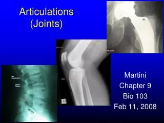
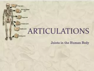
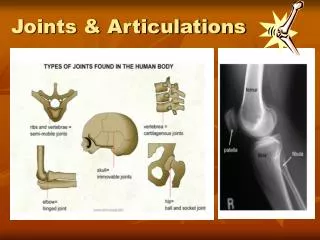
Articulations (Joints). Connect 2 bones. Classified by structure or function. Classification by structure 1. Fibrous joints - bones held together by fibrous connective tissue 2. Cartilagenous joints - bones held together by cartilage 3. Synovial joints

E N D
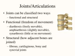
Connect 2 bones. • Classified by structure or function
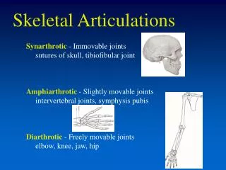
Classification by structure 1. Fibrous joints - bones held together by fibrous connective tissue 2. Cartilagenous joints - bones held together by cartilage 3. Synovial joints - complex structure with cartilage and cavities Classification by function 1. Synarthroses Immovable joints 2. Amphiarthrose Slightly movable joints 3. Diarthroses Freely movable joints Joint classification
General background • 1 extra joint category Bony joint (synostosis) • Immobile (immovable) • Fond when 2 bones ossify together (essentially merge or weld together) • Some cranial sutures (fissures in the cranium) become ossified • When the growth plate fuses/calcifies over (joining the epiphysis and diaphysis in long bones)
Fibrous joint (synarthrosis) • 1.Suture • 2. Gomphosis • 3. Syndesmosis
Suture: immobile joints in the skull (the fissures you see) through the thickness of the outer compact bone -“serrate”= wavy lines (like a serrated knife) - skull: parieta/frontal bones “lap”/ “squamous”= diagonal line Skull: temporal/parietal bones “plane” / “butt”= straight line Paired maxillary and palatine bones 1. Sutures
2. Gomphosis • where teeth attach to bone • Tooth is “held in place” by fibrous peridontal ligament” made of collagen that originates from the jaw bone • Fibrous “joint” permits slight movement when biting • An important sensory mechanism that lets you know how hard or soft you are biting
Bacterial infection destroys the ligament holding the teeth in place Promoted by the presence of plaque Gingivitis (gum disease)
A chronic lack of vitamin C leads to scurvy Vitamin C is necessary for collagen synthesis With decreased collagen synthesis, joints are weakened, wounds do not heal well. How do you treat scurvy? Scurvy
3. Syndesmosis • Colllagen fibers bind 2 bones • More mobility than suture or gomphosis, but still very limited • Tibia – fibula at distal end • Radius – ulna at distal ends
Tibia/fibula Radius/ulna Note how far apart the radius/ulna are from one another compared to the tibia/fibula. This is one reason why your forearm is more “mobile” or flexible than your lower leg.
Synchondoses: when bone is bound /joined byhyaline cartilage The growth plate in long bones The first rib attaches to the sternum (ONLY the first rib) Symphyses: where bones are joined by fibrous cartilage Pubic bone (pelvis) Between vertebrae Cartilaginous joints: (amphiarthrosis)
Trick question • How/why do infants and children have more joints than adults?
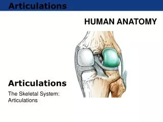
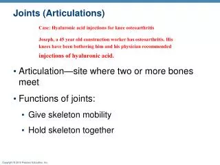
- Very complex “synovial” joint because the joints are separated by a space (“synovial cavity”), where synovial fluid is retained Synovial fluid rich in albumin (blood protein) and hyaluronic acid (a lubricant) Synovial joints
Synovial joints • Joint membrane: • Outer fibrous capsule (continuous with the periosteum) • Inner synovial membrane secretes the synovial fluid into the joint capsule • Each bone involved in the joint is covered by articular cartilage 2 mm thick -The bones making up the joint are “held together” by ligaments • Help to locate the bones in the correct “place” so that their articular cartilage “rides” correctly
Ball & Socketjoints (hip and shoulder) Hinge joints (ulna and femur…knee, fingers and toes) Pivot joints (radius and ulna…forearm/elbow and the first 2 vertebrae of your spine) Saddle joints (thumb, sternum/clavicle) Condyloid joints (fingers) Types of joints: classification by shape
Ball & Socketjoints (hip and shoulder) The ONLY multi-directional joints you have Types of joints: classification by shape
Ball & Socket joints Hinge joints (ulna and femur…knee, fingers and toes) 1 range of movement (flip-phone) Types of joints: classification by shape
Ball & Socket Hinge joints Pivot joints (radius and ulna…forearm/elbow and the first 2 vertebrae of your spine) When 1 bone spins on another, when you say no, or turn a door knob) Types of joints: classification by shape
Ball & Socketjoints Hinge joints Pivot joints Saddle joints (thumb, sternum/clavicle) 2 ranges of motion (hold something tight = sagittal plane, spread hand = frontal plane) Condyloid joints (fingers) 2 ranges of motion: make a fist = sagittal plane, spread fingers = frontal plane Types of joints: classification by shape
Ball & Socketjoints Hinge joints Pivot joints Saddle joints Condyloid joints Gliding joints Bone surface almost flat Bones slide along each other Ex: carpal bone, patella-femur Types of joints: classification by shape
Complex joints Many joints have characteristics of multiple joint types • Elbow is both a hinge joint (where ulna and humerus meet), as well as a pivot joint (where radius and ulna meet) • Knee is similar to the elbow joint, without as much flexibility (much stronger and limited) • Temporo-mandibular joint has both lateral and dorsal/ventral movement
In a few joints (jaw = temporo-mandibular), (sternum/collarbone =sterno-clavicular), (ankle = distal tibia-fibula), there is an articular disc In your knee, this is called a meniscus for guidance of the knee bones for shock absorption Complex synovial joints
Other structures present in synovial joints: Tendons: collagen-based connective tissue that hold muscle to bone Ligaments: collagen-based connective tissue that hold bone to bone Bursa: fibrous “sac” that holds synovial fluid Wraps around muscles prevent rubbing “bursitis” = inflammation of the bursa “tendon sheath” Accessories to synovial joints
Movements of various joints • Your shoulder (ball and socket) is a good example of a joint that is “multiarticulate” • The ball and socket allows for: • Abduction of the arm (flap your arms like a bird, jumping jacks) • Flexion of the arm (extending to shake a hand or open a door) • Rotation of the arm (sweeping your desk)
Movements of various joints Abduction = raising arms above head (moving away from the anatomical position in the FRONTAL PLANE) Adduction = returning arms to side (returning to anatomical position in the FRONTAL PLANE) The terms abduction and adduction refer to the ACTION, NOT THE APPENDAGE that you are moving (you can also abduct and adduct your legs at the hip joint).
Movements of various joints Elevation = lifting body part in the FRONTAL PLANE (moving away from the anatomical position) In this case, the model is raising their shoulders in the frontal or coronal plane Depression = returning to anatomical position from elevation These terms refer to movement FROM the anatomical position, as well as back TO the anatomical position. These movement terms also refer to the PLANE of movement. In this case, we are dealing with the FRONTAL or CORONAL plane.
Movements of various joints Protraction = moving body part forward (anterior movement) in a transverse or horizontal direction Retraction= posterior movement (“pushing out” his chest…he’s really pulling back his shoulders”) With these terms, there is reference to direction and plane of movement, however, this DOES NOT refer to the movement FROM or TO the anatomical position.
Movements of various joints Flexion = decreasing joint angle In this case, the models elbow is in flexion Extension= increasing joint angle to “zero position” (where it would “naturally rest) Hyperextension= moving beyond the zero position Note that this movement DOES NOT refer to the plane of a joint, is only refers to the ANGLE of the joint.
Movements of various joints Hyperextension= moving beyond the zero position
Movements of various joints Circumduction = 1 end of the joint remains stationary, the other end makes a CIRCLE. Rotation = turning a joint longitudinally on 1 bone of the joint
Movements of various joints Supination =ONLY for FOREARM, when you turn your forearm so that the palms face forward or ventral (anatomical position) Pronation= ONLY for the forearm, when you turn your forearm so that the palm faces posterior or dorsal
Movements of hand joints Radial flexion= tilting the hand towards the thumb (towards the radius) Ulnar flexion= tilting hand towards the pinky finger (towards the ulna) Abductionof theFINGERSis when you spread your fingers apart In terms of yourfingers: Flexion= curling fingers (like a fist) Extension= pointing your fingers
Movements of hand joints Your THUMB is turned 90 from the rest of your fingers, so the previous “terms” for finger movement are DIFFERENT for your thumb Abduction= bring your thumb to your index finger (the “OK” sign) Opposition= touch pinky with thumb Reposition= return to “zero” from the opposition position In terms of yourTHUMB: Flexion= bending towards the palm of your hand Extension= pointing thumb up (hitch-hiking) Hyperextension= when you make a 90 angle between your thumb and index finger (straight up hitch hiker thumb)
Movements of foot/ankle joints In terms ofANKLEmovements Dorsiflection= lift toes up (standing on your heels) Zero position= feet flat on the ground, standing up Plantar flexion= pointing toes down (standing on your toes) In terms of yourFEET: Your ankle can “roll” just like your wrist can “wave” Inversion= turning the plantar surface towards the median (standing on the outside of your feet) Eversion= turning the plantar surface laterally (standing on the arches of your feet)
The scapula is connected to the thorax only by the clavicle (collar bone) Connection at the acromion process (look for a sharp hook) The joint between the clavicle and acromion = acromial-clavicular joint Complex joint: humero-scapular joint (shoulder)
Humero-scapular joint Posterior view Anterior view Note how the ligaments and tendons wrap around the joint to hold the head of the humerus into the glenoid cavity. Also note how shallow the “socket” on the scapula is; an effort to increase mobility of this joint
Loose ball and socket joint. More mobility Muscles and ligaments stabilize the joint
Actually 2 joints in 1 area Humero-ulnar joint Humero-radial joint Both enclosed by 1 single synovial joint capsule 3rd joint in the elbow: radio-ulnar joint head of the radius - radial notch in the ulnus important because it permits the forearm rotation (supine/prone) rotation Your tibia/fibula (shins) cannot rotate as well Complex joint: Elbow joint
Features of the hand and wrist Also remember that your hand and wrist comprise a number of different joint types: saddle, gliding and condyloid. Together, these joints give your hands and fingers a great deal of and mobility
Rheumatoid arthritis - An autoimmune disease
Osteoarthritis - Bone degeneration due to old age
Accumulation of synovial fluid in the bursa – due to chronic or acute irritation An extreme case! Bursitis
Sprain: ligament tear Strain: Tendon or muscle tear Sprain and Strain