Download
1 / 26
260 likes | 548 Views
Disclosure. No Relevant Financial Relationships with Commercial InterestsI will not reference an unlabeled or unapproved use of a drug or product in my presentation Marzouq Qubti, MDRheumatology Rounds 9-12-2008 . . Objectives. To Consider the Broad range of conditions that contribute to MeningitisTo Consider Rheumatic etiologies during the evaluation of meningitis.

E N D
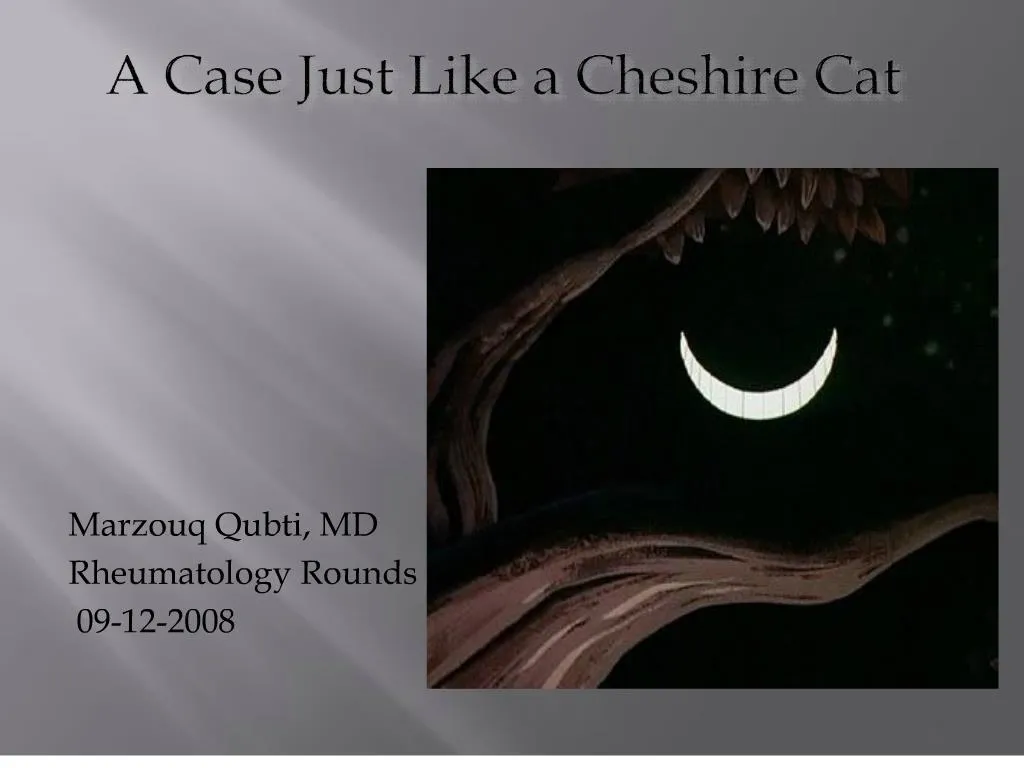
1. A Case Just Like a Cheshire Cat
Marzouq Qubti, MD
Rheumatology Rounds
09-12-2008 Welcome to the first academic Rounds of the year. This is a thought provoking case which we were consulted on on Wednesday August 13th 2008 with Dr. gelber.Welcome to the first academic Rounds of the year. This is a thought provoking case which we were consulted on on Wednesday August 13th 2008 with Dr. gelber.
2. Disclosure No Relevant Financial Relationships with Commercial Interests
I will not reference an unlabeled or unapproved use of a drug or product in my presentation
Marzouq Qubti, MD
Rheumatology Rounds
9-12-2008
3. Objectives To Consider the Broad range of conditions that contribute to Meningitis
To Consider Rheumatic etiologies during the evaluation of meningitis
4. Consult for FUO Consult 8-13-2008
34 Year old female transferred from OSH after over three weeks of inpatient hospitalization for intermittent fevers.
5. HPI 34 Year old Asian female lawyer, mother of two.
3 weeks of fever despite inpatient evaluation
Healthy until 7-7-2008 when she developed Flu-like symptoms
She was 8 months pregnant at the time
Treated for presumed UTI
34 year old female of Asian background who was well until her 8th month of gestation right after the fourth of July weekend when she developed malaise and maylagias. She saw her PBGYN on 7-9-2008 and was treated with cephlexin for a presumed UTI with a UA eventually coming back negative.34 year old female of Asian background who was well until her 8th month of gestation right after the fourth of July weekend when she developed malaise and maylagias. She saw her PBGYN on 7-9-2008 and was treated with cephlexin for a presumed UTI with a UA eventually coming back negative.
6. Timeline � July 2008 7-7-2008 : Flu Like Symptoms
7-10-2008: Worsening Symptoms
7-12-2008---7-14-2008: Improvement
7-17-2008: Admission #1
Temp 103
CSF 1,500 WBC
Gluc 23
Protein 182
7-19-2008: Ten Day Treatment course
7-22-2008: C-Section
7-28-2008: Hives �red dots� � stopped amoxicillin
7-31-2008: Discharged
60% N
30% Lymphs
10% monos
Treatment with Ampicillin, Ceftriaxone, and vancomycin along with decacdron
She received 4 days of decardon ampicillin stopped due to lower extremity maculo papular rash.
The patient reports improvement between 7-12-08 and 7-14-08 to the point where was able to go to work and do yoga.
She told us she was never back to normal 100% but enough to go to work and do yoga
C-Section because of concerns on the baby. Mainly a change in the babies acceleration.
After decadron for 4 days- van and ceftriaxone for ten days and amox for nine days, she was DC�d home
When indicated, dexamethasone is given 15 to 20 minutes before or at the time of antibiotic administration. Two dose regimens have shown efficacy: 0.15 mg/kg every six hours for four days in the European trial and 0.4 mg/kg every 12 hours for four days in the Vietnamese trial.
For adults in the developed world with known or suspected pneumococcal meningitis and a Glasgow coma score between 8 and 11, we suggest administration of dexamethasone (Grade 2B). For all other adults in the developed world with confirmed or suspected bacterial meningitis, we suggest not administering dexamethasone (Grade 2B). (See "Developed regions" above).
For adults from areas of the developing world with high HIV prevalence and known or suspected bacterial meningitis, we recommend not administering dexamethasone (Grade 1B). (See "Developing regions" above).
7. Differential Diagnosis
8. Time course: August 2008 8-4-2008: Fever recurrence- 100 � F
Intensity of rash on legs correlates with fevers
ED visit: ID Consult: Drug Fever
+ Sore Throat.
8-7-2008: Fevers 103�
Admission #2
CSF : WBC: 480. 62% lymphocytes
Protein 206
Glucose 34
Brain MRI/CT/CSF PCR AFB �
8-11-2008: JHH Transfer
37% N-62% lymphocytes
This was diagnosed as a delayed drug fever one week after stopping the amoxicillin37% N-62% lymphocytes
This was diagnosed as a delayed drug fever one week after stopping the amoxicillin
9. PMHx
Prior Positive PPD
No BCG
No known exposure
Arthralgias
3 years old-8 years old
Nondescript bilateral hand stiffness and pain intermittently
She reported no treatment for her positive PPD
Arhtrlagias were in her bilateral knees. She used to cry at night from the pain
She did not seek medical attention and the pain had gotten so bad that she was sent to an acupuncturist
For itShe reported no treatment for her positive PPD
Arhtrlagias were in her bilateral knees. She used to cry at night from the pain
She did not seek medical attention and the pain had gotten so bad that she was sent to an acupuncturist
For it
10. Family History: Brother has intermittent knee pain
Grandmother had an arthritis
Healthy 3 year old daughter
Healthy newborn daughter
11. Social history: Non-smoker
No IVDU
No cocaine
No Tattoos
Two Units of Packed Red Blood Cells
Born in South Korea. Moved to US at the age of 6.
Lives in DC
Visited Korea, China, Taiwan, Japan, Italy, and Cambodia in the past 10 years. During the c-section delivery
She used to live in a suburban area of DC where mold was present in the old house
IN their current neighborhood , the patient did report rodents on the streets nearby but not inside the buildingDuring the c-section delivery
She used to live in a suburban area of DC where mold was present in the old house
IN their current neighborhood , the patient did report rodents on the streets nearby but not inside the building
12. ROS Intermittent Headache
Night Sweats
Intermittent Arthralgias.
She denied ever taking Ampicillin prior to Outside hospitalization ROS: When we saw the patient on 8-13-2008ROS: When we saw the patient on 8-13-2008
13. Physical Examination 105� : Tmax 110-120: Pulse when febrile
CV: 2/6 Systolic flow murmur 2nd intercostal
space
Abdomen: low transverse laparotomy scar
No rashes.
14. Differential DiagnosisPost Second Hospitalization
15. Na 138
K 4.2
Cl 104
BUN 9
Gluc 113
Calcium 9.3
T Bili 0.3
Alk Phos 103
Peripheral blood smear Burrs and ovalocytes and fragmentsNa 138
K 4.2
Cl 104
BUN 9
Gluc 113
Calcium 9.3
T Bili 0.3
Alk Phos 103
Peripheral blood smear Burrs and ovalocytes and fragments
17.
CSF IGG 8.5
CSf Albumin 59
Serum IgG 591- low normal being 751-1560
No oligoclonal bands
Cryptococcal antigen csf NEGATIVE INDIA INK STAIN CSF NEGATIVE
1) By the age of 30, 80% of the population is EBV infected
2)By the age of 6, 60% of the population is CMV infected
3)Epidemiologically, Primary CMV/EBV at 34 is rare.
4)A primary Infection would usually be associated with a negative IgG (Both IgGs positive)
5)CMV/EBV are chronic, latent infections that can occasionally replicate at subclinical levels and induce transient IgM spikes
6)CMV PCR at Outside hospital was negative
) False + PCR in CSF for EBV and CMV theoretically possible in the setting of any brain inflammatory process (Latent virus in lymphocytes). Hence viral culture is more specific for active CMV and EBV
8) Her baby does not have TORCH syndrome
LCM- Human zoonoziz causes by rodent borne areanavirus urine and feces of rodents- including mice and rates and hamsters= influenza like symptoms headache and menignismus- a minortiy will develop orchitis , parotitis, myoperciarditis or arthritis
Aseptic meningitis is the most frequent extraslaivary complication of mumps virus infection
White count in cryptococcal meningitis less than 50 with a mononuclear predominanceCSF IGG 8.5
CSf Albumin 59
Serum IgG 591- low normal being 751-1560
No oligoclonal bands
Cryptococcal antigen csf NEGATIVE INDIA INK STAIN CSF NEGATIVE
1) By the age of 30, 80% of the population is EBV infected
2)By the age of 6, 60% of the population is CMV infected
3)Epidemiologically, Primary CMV/EBV at 34 is rare.
4)A primary Infection would usually be associated with a negative IgG (Both IgGs positive)
5)CMV/EBV are chronic, latent infections that can occasionally replicate at subclinical levels and induce transient IgM spikes
6)CMV PCR at Outside hospital was negative
) False + PCR in CSF for EBV and CMV theoretically possible in the setting of any brain inflammatory process (Latent virus in lymphocytes). Hence viral culture is more specific for active CMV and EBV
8) Her baby does not have TORCH syndrome
LCM- Human zoonoziz causes by rodent borne areanavirus urine and feces of rodents- including mice and rates and hamsters= influenza like symptoms headache and menignismus- a minortiy will develop orchitis , parotitis, myoperciarditis or arthritis
Aseptic meningitis is the most frequent extraslaivary complication of mumps virus infection
White count in cryptococcal meningitis less than 50 with a mononuclear predominance
19. RF and CMV/EBV Ig Peripheral Blood Mononuclear cells from normal controls but not RA patients appeared to be responsive to viral antigen stimulation (CMV and EBV) and produced RF. Immune response to CMV/EBV correlates with the presence of RF
Ferraro A.S. Newkirk M. Correlative studies of Rheumatoid factors and anti-viral antibodies in patients with rheumatoid arthritis Clin Exp Immunol 1993 ; 92:425-431
I put this slide up because I was curious if it was ever shown in the literature that RF is actually elevated in acute EBV/CMV and this actually documented production of RF from Mononuclear cells stimulated with CMV/EBV antigenI put this slide up because I was curious if it was ever shown in the literature that RF is actually elevated in acute EBV/CMV and this actually documented production of RF from Mononuclear cells stimulated with CMV/EBV antigen
20. Imaging MRI of the brain with Gadolinium
Done at OSH and Read at JHH
Dedicated MRA was not done
Major vascular flow noted to be normal
CT of the abdomen/Pelvis:
Prominent Uterus
�Butterfly vertebra of T10�
Left Lower Lobe 5 mm Granuloma
Echocardiogram: No vegetation Echo negative for any vegatation.Echo negative for any vegatation.
21. Butterfly vertebrae result from the failure of fusion of the lateral halves of the vertebral body because of persistent notochondal tissue between them
They occur most often in Bulldogs, Pugs, and Boston Terriers
Notochord: flexible rod shaped body found in embryosof all chordates. Cells derived from the mesodermand defines the primitive axis of the embryo. In lower vertrates, it persists as the main axial support while in higher vertebrates it is replaced by the vertebral columnThey occur most often in Bulldogs, Pugs, and Boston Terriers
Notochord: flexible rod shaped body found in embryosof all chordates. Cells derived from the mesodermand defines the primitive axis of the embryo. In lower vertrates, it persists as the main axial support while in higher vertebrates it is replaced by the vertebral column
22. Can we Give this patient a diagnosis Is this rheumatic or infectious ?
23.
Our Differential Diagnosis
24. MeningitisBacterial Neisseria meningitidis
Streptococcus pneumoniae
Listeria monocytogenes
Coagulase negative Staphylococcus
Staphylococcus Aureus
GNR: Elderly more presdisposed
Haemophilus Influenzae
N.Meningitides: Site of entry Nasopharynx: all ages: at times complement deficiency may be a cause of this
S.pneumo: Nasopharynx or direct extension across skull fracture all ages
L.Monocytogenes: GI tract: Placenta: All ages: defect in cell mediated immunity can cause it
Coag negative staph: Dermal skin: foreign body: All ages susceptible: predisposing condition surgery or shunt
S. Aureus: Dermal bactermeia or forign body predisposing condition: endocarditis, surgery, ventrucular chunt
Hflu: Through the nasopharynx: infants and children if not vaccinated: Diminsihed humoral immunity
1/3 patients will not present with fever nuchal rigditiy and change in mental status
Pregnant women 20 times more likely to get listeria than non pregnant women
Recs are not to eat luncheon meats, deli meats, unless they are reheated to be steaming hot- pasterization kills and therefore you should avoid unpasteurized cheese N.Meningitides: Site of entry Nasopharynx: all ages: at times complement deficiency may be a cause of this
S.pneumo: Nasopharynx or direct extension across skull fracture all ages
L.Monocytogenes: GI tract: Placenta: All ages: defect in cell mediated immunity can cause it
Coag negative staph: Dermal skin: foreign body: All ages susceptible: predisposing condition surgery or shunt
S. Aureus: Dermal bactermeia or forign body predisposing condition: endocarditis, surgery, ventrucular chunt
Hflu: Through the nasopharynx: infants and children if not vaccinated: Diminsihed humoral immunity
1/3 patients will not present with fever nuchal rigditiy and change in mental status
Pregnant women 20 times more likely to get listeria than non pregnant women
Recs are not to eat luncheon meats, deli meats, unless they are reheated to be steaming hot- pasterization kills and therefore you should avoid unpasteurized cheese
25. Aseptic Meningitis Definition Symptom complex that is produced by any one of numerous infective agents, the majority of which are viral ( but a few are bacterial �mycoplasma, Q fever, other rickettsial infections, etc.)
May be some minor changes to the brain, but insufficient in severity to cause alteration on CT or MR.
Ropper A
Principles of Neurology 8th Edition 2005
26. Aseptic MeningitisDefinition Brudzinski and Kernig help very little in Aseptic meningitis
Originally tested for severe late stage meningitis
Jolt accentuation of headache had a sensitivity of 97 percent and a specificity of 60 percent for the diagnosis of CSF pleocytosis
Thomas KE Hasbun R Jeckel R. The diagnostic accuracy of Kernig�s sign, brudzinski�s sign, and nuchal rigidity in adults with suspected meningitis.Clin Infect Dis 2002 Jul 1; 35:46-52
Jolt accentuation of headache: the most sensitive sign of CSF pleocytosis. AU Uchihara T; Tsukagoshi H SO Headache 1991 Mar;31(3):167-71.
Clues on physical exam- a diffuse maculopapular exanthem in a mildly ill patient may be consistent with an enteroviral infection, primary HIV, or syphilis
Parotitis suggest mumps
Classic Bruzinski: Spontaneous flexioun of the hips during attempted passive flexion of the necy
Kernig: inability or reluctance to extend knee when the hip is flexed at 90 degrees
297 adults suspected to have meningitis were prospectively evlauted for these meningeal signsbefore lumbar puncture was done Sensitivty to have positive Kernig was 5% and Brudz 5%Nuchal rigidity is about 30%. The diagnostic accuracy was not significantly better in patients with moderate meningeal inflammation (over a 100 WBC)
Among 34 patients with pleocytosis, 33 had jolt accentuation (sensitivity: 97.1%), while only 5 of them had neck stiffness or Kernig's sign. Among 20 patients without pleocytosis, 12 had no jolt accentuation (specificity: 60%). We found jolt accentuation to be the most sensitive sign of CSF pleocytosis. If jolt accentuation is noted in a febrile patient associated with recent onset headache, the CSF should be examined even in the absence of neck stiffness or Kernig's sign. �Clues on physical exam- a diffuse maculopapular exanthem in a mildly ill patient may be consistent with an enteroviral infection, primary HIV, or syphilis
Parotitis suggest mumps
Classic Bruzinski: Spontaneous flexioun of the hips during attempted passive flexion of the necy
Kernig: inability or reluctance to extend knee when the hip is flexed at 90 degrees
297 adults suspected to have meningitis were prospectively evlauted for these meningeal signsbefore lumbar puncture was done Sensitivty to have positive Kernig was 5% and Brudz 5%Nuchal rigidity is about 30%. The diagnostic accuracy was not significantly better in patients with moderate meningeal inflammation (over a 100 WBC)
Among 34 patients with pleocytosis, 33 had jolt accentuation (sensitivity: 97.1%), while only 5 of them had neck stiffness or Kernig's sign. Among 20 patients without pleocytosis, 12 had no jolt accentuation (specificity: 60%). We found jolt accentuation to be the most sensitive sign of CSF pleocytosis. If jolt accentuation is noted in a febrile patient associated with recent onset headache, the CSF should be examined even in the absence of neck stiffness or Kernig's sign. �
27. Aseptic Meningitis In order of frequency:
1)Enteroviruses: echoviruses and coxsackie
2)Mumps
3)HSV-2
4)Lymphocytic choriomeningitis
5)EBV, CMV, HSV-1, HIV leptospirosis, and M. pneumoniae, lyme borreliosis
6)Influenza, adenoviruses.
7)Anthropod borne viruses
Ropper, Brown
Principles of Neurology, 8th Edition
Enteroviruses: Summer or fall time is entrovirus Coxsachivirus, Echovirus, non polio entrovirsues
HSV-2 : More common than HSV1 for meningitis. For encephalitis , it is HSV 1 (Almost exclusively)
Mycoplasma, lyme , rickettsial infections are considered aseptic in nature
The syndrome
Lymphocytic pleocytosis
Headache meningeal irritation
Fever , lethargy , stupor confusion, irritability,
Normal CSF glucose
Mainly mononuclear pleocytosis except the first few days where there is a predominance of neutrophils
Small or variable increase in protein
AS A RULE THE GLUCOSE CONTENT SHOULD BE NORMAL- LOW SUGAR WITH A LYMPHOCYTIC PLEOCYTOSIS MAKES YOU THINK OF FUNGAL OR TUBERCULOUS, CANCER, OR SARCOID
28. Drugs and Aseptic meningitis
Bai , glass: Resident and Staff Physician: 2005D
Mechanisms to explain Drug Induced meningitis:
1)Delayed Hypersensitivity reaction
2)Direct meningeal irrtiation
CSF usually presents as a neutrophilic pleocytosisMechanisms to explain Drug Induced meningitis:
1)Delayed Hypersensitivity reaction
2)Direct meningeal irrtiation
CSF usually presents as a neutrophilic pleocytosis
29. Aseptic Meningitis-Other Foci of Bacterial infection near meninges
Partially treated bacterial Meningitis
Fungal, tuberculous meningitis, parasites
Neoplastic invasion
Lymphomatous
Carcinomatous 1)Iridocyclitis
Depgimentation of the hair strand and skin around the eyes
Loss of eyelashes
Syacusis and deafness
2) Follow bouts of general herpes. Anti-virals met
With little success. Fever headaches in two cycles for a few months
Intracranial epidermoid cysts in the brain can be a non infectious cause of Mollaret�s meningitis1)Iridocyclitis
Depgimentation of the hair strand and skin around the eyes
Loss of eyelashes
Syacusis and deafness
2) Follow bouts of general herpes. Anti-virals met
With little success. Fever headaches in two cycles for a few months
Intracranial epidermoid cysts in the brain can be a non infectious cause of Mollaret�s meningitis
30. Aseptic MeningitisRheumatic Sarcoidosis
Behcets
Sjogren�s Syndrome
RA
Wegener�s Granulomatosis
Primary Angiitis of the CNS
SLE
Sarcoidosis: Neurologic Complications can occur in 5 % of patients with sarcoid- central and peripheral nerve involvement. Peripheral Facial Nerve palsy is common, seizures,
Peripheral facial nerve palsy develops in over 50 percent of patients with neurosarcoidosis [2] . The facial nerve palsy can be unilateral or bilateral (simultaneous or sequential) and recurrent. Optic neuropathy and cranial nerve VIII dysfunction can lead to intermittent or progressive visual, auditory, or vestibular dysfunction.
Prospective studies suggest that from 50 to 78 percent of neurologic episodes are caused by secondary factors [2,8] , including:
Infections associated with immunosuppressive therapy
Metabolic complications of other organ system failure, such as uremia
Hypertension
Toxic effects of therapy (particularly corticosteroids)
RA
Rheumatoid pachymeningitis is a rare CNS complication of rheumatoid arthritis (RA). This occurs in patients with long-standing seropositive RA with extra-articular involvement (ExRA). We reviewed the literature searching PubMed from 1971 to present using terms the "rheumatoid arthritis" and "pachymeningitis." References from identified articles were also selected. Only 20 histopathologically proven cases have been reported. The prevalence of this disorder is unknown. We describe two patients who met American College of Rheumatology criteria for RA, with biopsy-proven pachymeningitis.
Neurological involvement in WG is uncommon at the onset of disease1,2, but may develop in 22% to 54% of cases over time1,2,4-6. The most frequent nervous system manifestation is peripheral neuropathy, which occurs in 15% to 43% of cases1,3,5,7. The most common form of peripheral neuropathy is mononeuritis multiplex, followed by distal symmetric sensorimotor polyneuropathy. Involvement of the central nervous system (CNS) is infrequent. Among 249 cases of WG reported by Anderson, et al, there were 12 patients with CNS vasculitis, and 32 had evidence of CNS granuloma6. CNS involvement occurred in 8% of 158 patients followed at the US National Institutes of Health1 and in 11% in a more recent European series3. The spectrum of CNS involvement includes stroke, cranial nerve abnormalities, cerebrovascular events, seizures, and meningeal involvement.
Meningeal involvement, illustrated in our patient by contiguous pachymeningitis and leptomeningitis, is rare in WG. Among 324 consecutive patients seen at the Mayo Clinic there were only 2 reported cases of meningeal involvement5. Drachman found 7% of 104 patients
Headache is almost always the first symptom of meningeal involvement in WG. Later in the course of the disease other abnormalities may develop. Among them cranial nerve palsy, seizures and encephalopathy are the most frequent. Diagnosis is obtained by neuroimaging, which may disclose two distinct patterns of meningeal thickening: diffuse or focal. 62.9% of patients tests positive for ANCA. Histology typically shows necrotizing granulomatosis. Meningeal involvement is by far more frequent in the setting of localized WG. Meningitis is a rare complication of WG. It usually develops in patients with localized disease who are more likely to have destructive lesions of the upper airways. It may be recognized by a constellation of clinical and radiological findings and by histological signs of necrotizing granulomatosis, with little or no vasculitis.
PMID: 16859598 [PubMed - indexed for MEDLINE]
Focal parenchymal lesions and complications of vascular thrombosis are the most common abnormalities-BEHCETSSarcoidosis: Neurologic Complications can occur in 5 % of patients with sarcoid- central and peripheral nerve involvement. Peripheral Facial Nerve palsy is common, seizures,
Peripheral facial nerve palsy develops in over 50 percent of patients with neurosarcoidosis [2] . The facial nerve palsy can be unilateral or bilateral (simultaneous or sequential) and recurrent. Optic neuropathy and cranial nerve VIII dysfunction can lead to intermittent or progressive visual, auditory, or vestibular dysfunction.
Prospective studies suggest that from 50 to 78 percent of neurologic episodes are caused by secondary factors [2,8] , including:
Infections associated with immunosuppressive therapy
Metabolic complications of other organ system failure, such as uremia
Hypertension
Toxic effects of therapy (particularly corticosteroids)
RA
Rheumatoid pachymeningitis is a rare CNS complication of rheumatoid arthritis (RA). This occurs in patients with long-standing seropositive RA with extra-articular involvement (ExRA). We reviewed the literature searching PubMed from 1971 to present using terms the "rheumatoid arthritis" and "pachymeningitis." References from identified articles were also selected. Only 20 histopathologically proven cases have been reported. The prevalence of this disorder is unknown. We describe two patients who met American College of Rheumatology criteria for RA, with biopsy-proven pachymeningitis.
Neurological involvement in WG is uncommon at the onset of disease1,2, but may develop in 22% to 54% of cases over time1,2,4-6. The most frequent nervous system manifestation is peripheral neuropathy, which occurs in 15% to 43% of cases1,3,5,7. The most common form of peripheral neuropathy is mononeuritis multiplex, followed by distal symmetric sensorimotor polyneuropathy. Involvement of the central nervous system (CNS) is infrequent. Among 249 cases of WG reported by Anderson, et al, there were 12 patients with CNS vasculitis, and 32 had evidence of CNS granuloma6. CNS involvement occurred in 8% of 158 patients followed at the US National Institutes of Health1 and in 11% in a more recent European series3. The spectrum of CNS involvement includes stroke, cranial nerve abnormalities, cerebrovascular events, seizures, and meningeal involvement.
Meningeal involvement, illustrated in our patient by contiguous pachymeningitis and leptomeningitis, is rare in WG. Among 324 consecutive patients seen at the Mayo Clinic there were only 2 reported cases of meningeal involvement5. Drachman found 7% of 104 patients
Headache is almost always the first symptom of meningeal involvement in WG. Later in the course of the disease other abnormalities may develop. Among them cranial nerve palsy, seizures and encephalopathy are the most frequent. Diagnosis is obtained by neuroimaging, which may disclose two distinct patterns of meningeal thickening: diffuse or focal. 62.9% of patients tests positive for ANCA. Histology typically shows necrotizing granulomatosis. Meningeal involvement is by far more frequent in the setting of localized WG. Meningitis is a rare complication of WG. It usually develops in patients with localized disease who are more likely to have destructive lesions of the upper airways. It may be recognized by a constellation of clinical and radiological findings and by histological signs of necrotizing granulomatosis, with little or no vasculitis.
PMID: 16859598 [PubMed - indexed for MEDLINE]
Focal parenchymal lesions and complications of vascular thrombosis are the most common abnormalities-BEHCETS
31. Adult Onset Still�s DiseaseYamagushi Criteria Major Criteria:
Fever of at least 39 degrees lasting at least 1 week
Arthralgias or arthritis lasting 2 weeks
Nonpruritic macular rash that is salmon colored
Leukocytosis (10,000/MicroL), 80% Granulocytes
Etiology of Still�s unknown bacterial suspicion yersinia- mycoplasma
HLA B-17 associationEtiology of Still�s unknown bacterial suspicion yersinia- mycoplasma
HLA B-17 association
32. Adult Onset Still�s DiseaseYamaguchi Criteria Minor Criteria:
Sore Throat
Lymphadenopathy
Hepatomegaly
Abnormal Liver Function Studies
Negative ANA and RF
Yamaguchi M; Ohta A. Preliminary criteria for classification of Adult Still�s disease. J Rheumatol 1992; 19; 424-30
The presence of any infection malignancy or other rheumatic disorder known to mimic ASD precludes the diagnosis
Etiology of Still�s unknown bacterial suspicion yersinia- mycoplasma
HLA B-17 associationThe presence of any infection malignancy or other rheumatic disorder known to mimic ASD precludes the diagnosis
Etiology of Still�s unknown bacterial suspicion yersinia- mycoplasma
HLA B-17 association
33. E.G.L Bywaters E. G. Bywaters: Still's disease in the adult.Annals of the Rheumatic Diseases, March 1971, 30 (2): 121-133
Bywaters EG. The Cheshire cat syndrome. Postgrad Med J. 1968; 44: 19�22. As a Gelber, MD Digression, Bywaters was the first to explain the pathophysiology of ARF after crush injuries and rhabdo when he reported the delayed death after rescue of four English victims after the London Blitz in 1941. he names the syndrom the Bywaters syndrome but that did not catch on. As a Gelber, MD Digression, Bywaters was the first to explain the pathophysiology of ARF after crush injuries and rhabdo when he reported the delayed death after rescue of four English victims after the London Blitz in 1941. he names the syndrom the Bywaters syndrome but that did not catch on.
34. Once a joint is involved , recurrence is not infrequent.
Seldom incapacitating
Knees, wrists, fingers frequently affectedOnce a joint is involved , recurrence is not infrequent.
Seldom incapacitating
Knees, wrists, fingers frequently affected
35. Our Patient�s Prognosis
Elkon K.B , Hughes G.R.V, Bywaters, E.G.L. Adult Onset Still�s Disease Twenty year follow-up and Further Studies of Patients with Active Disease. Arthritis and Rheumatism . 1982 25; 647-5410 Fever rash arthritis most common symptoms during these exacerbations Fever rash arthritis most common symptoms during these exacerbations
36. Adult Onset Still�s DiseaseOriginal Rash Description �All Fourteen patients showed a typical Still�s rash characterized by small macules often with perimacular pallor due to deviation of arteriolar blood from the surrounding skin �.The macules come up with fever , usually therefore towards 6pm and may be seen outlining friction lines. Occasionally they may be slightly raised, only very rarely itchy. Usually on the limbs, the rash may also appear on the face and trunk�
37. Evanescent Rash Examine the patient in the evening with the fever spike
33% of patients can have the rash precipitated by rubbing
(Koebner�s phenomenon)
www.stillsdisease.org
38. Evanescent Rash
www.stillsdisease.org
39. Adult Onset Still�s DiseaseCNS Manifestations Original 14 patients described by Bywaters had no CNS involvement.
40. The CNS cases here were all reported before any of the aseptic meningitis cases reported in the next slide.The CNS cases here were all reported before any of the aseptic meningitis cases reported in the next slide.
41. Spinal Taps on Patient JI.
43. The Cheshire Cat Syndrome
44. In 1968, Bywaters diagnosed three patients, including a 6-year-old boy, with a vasculitis-like disorder that he assumed was most probably a partial variant of polyarteritis nodosa. However, autopsy study revealed none of the characteristic lesions.
In his report, Bywaters discussed the dilemma of administering treatment to patients who fail to fulfill all the diagnostic criteria of suspected diseases that, like the Cheshire cat, may develop gradually.
Bilavsky E et al. Literature Names for Pediatric Medical Conditions. Acta Pediatrica 2007 96:975-978
I thought it was interesting to note that the rheumatologist who eventually described AOSD has also philosophized on a medical conflict in which we have been faced with while dealing with the original diagnosis of his characterization.
This to some may sound anarchist but I feel , in our field, we deal with this all the time. He seemed convinced that had his patients stayed aive long they would have fulfilled the diagnostic criteriaI thought it was interesting to note that the rheumatologist who eventually described AOSD has also philosophized on a medical conflict in which we have been faced with while dealing with the original diagnosis of his characterization.
This to some may sound anarchist but I feel , in our field, we deal with this all the time. He seemed convinced that had his patients stayed aive long they would have fulfilled the diagnostic criteria
45. �Do these patients have Still�s disease or do they fit more easily into the pigeonholes constructed for adult disease, such as seronegative RA, AS, UC, arthritis without colitis, Psoriatic arthritis without psroiasis, or other varieties of the Cheshire Cat Syndrome ?�
E. G. Bywaters: Still's disease in the adult.Annals of the Rheumatic Diseases, March 1971, 30 (2): 121-133
Every paper you read on this guy, he is cross referencing himself. Here he is saying : � do these patients have this AOSD entity or do they have another better defined condition without fully declaring themselves to be that better defined condition�. I ask about our case: � Does Ms JI have idiopathic Aseptic meningitis due to a syndrome X or does she have AOSD that did not fully come out and declare himself�Every paper you read on this guy, he is cross referencing himself. Here he is saying : � do these patients have this AOSD entity or do they have another better defined condition without fully declaring themselves to be that better defined condition�. I ask about our case: � Does Ms JI have idiopathic Aseptic meningitis due to a syndrome X or does she have AOSD that did not fully come out and declare himself�
46. Treatment Options ?
47. Clinic Follow-up: 8-19-2008 Afebrile 97.7 � BP 88/65 (Her baseline!)
Negative Exam
Improved Energy. No headache or malaise
On Ibuprofen 400 mg QID ac and qhs standing
48. Follow up Clinic Visit : 9-2-2008 Ferritin 20
No Glycosylated Ferritin in lab at JHH
ESR 12
ANA now 1:160 from 1:80 nucleolar
Anti-DS DNA not Sent
49. AOSD and Pregnancy First case reported during pregnancy by Stein Et al in 1980
Nine Pregnancies reviewed retrospectively in small french Study- No relationship could be concluded
In Pregnant and post-partum Still�s, improved symptoms, recurrent flares, and stable disease all desrcibed
Flares noted in case reports after miscarriages and in the post partum setting Overall Post partum course trends towards worse outcomes. Patient with JRA with Atlanto-axial Subluxation and joint deformity--- difficult intubation s despite a higher rate for C-Section anyway due to hip prostehses and hip joint arthritisOverall Post partum course trends towards worse outcomes. Patient with JRA with Atlanto-axial Subluxation and joint deformity--- difficult intubation s despite a higher rate for C-Section anyway due to hip prostehses and hip joint arthritis
50. Summary Aseptic meningitis in the absence of positive cultures could yield a rheumatic cause especially if the course is sub-acute
Historically, Rheumatic diseases, like all ailments in general, have been known not to fulfill diagnostic criteria early in the course of their presentation.
A rheumatologic diagnosis may at times be challenged by seemingly discrepant clinical data (e.g. rheumatoid factor, positive viral IgM serology) that necessitates careful reconsideration of the working formulation
Adult Onset Still�s Disease can be a cause of aseptic meningitis in previously healthy adults
51.
�Sometimes I've believed as many as six impossible things before breakfast� Lewis Carroll
52. References: 1)Ohta A, Yamaguchi M: ASD: Review of 228 cases form the literature. J Rheumatol 14: 1139-46, 1987
2)Reginato A: AOSD: experience in 23 patients and literature review with emphasis on organ failure.17: 39-57, 1987
3)Ohta A, Yamaguchi M: ASD: A multicenter Survey of Japanese Patients. J Rheumatol 17: 1058-63, 1990
4)Denault A. Meningoencephalitis and peripheral neuropathy complicating ASD. Jrheumatol 17: 698-700, 1990
5)Fehmi, Malik et al :Neutrophilic Pleocytosis in CSF:AOSD. Internal Medicine. 42: 1039-41, 2003
6)Le Louet X:Still Disease in the adult and in pregnancy. Rev du Rhumatisime. 60(6):416-9
7)De Miguel E, Custa M et al. Adult Still�s and Pregnancy. J Rheumatol 1992;19:498
53. References 8)R Kumar, FC Guinto, Jr, JE Madewell, LE Swischuk, and R David.The Vertebral Body: Radiographic Configurations in Various Congenital and Acquired DisordersRadioGraphics 1988; 8: 455
9) Starosta M, Brandwein S. Clinical Manifestations and Treatment of Rheumatoid Pachymeningitis. Neurology 2007;68:1079-1080
10) Di Comite G et al. Clin Exp Rheumatology.Meningeal involvement in Wegener's granulomatosis is associated with localized disease. 2006 Mar-Apr;24(2 Suppl 41):S60-4.
11) Jolt accentuation of headache: the most sensitive sign of CSF pleocytosis. AU Uchihara T; Tsukagoshi H SO Headache 1991 Mar;31(3):167-71.