Download
1 / 22
410 likes | 1.16k Views
Protein Analysis. Prof. Conlon ’ s Lecture Unit 9 - Molecular Medicine 22 nd Feb 2003 Week 2. 4MedStudents.com. Why do we analyse proteins?.

E N D
Protein Analysis Prof. Conlon’s Lecture Unit 9 - Molecular Medicine 22nd Feb 2003 Week 2 4MedStudents.com
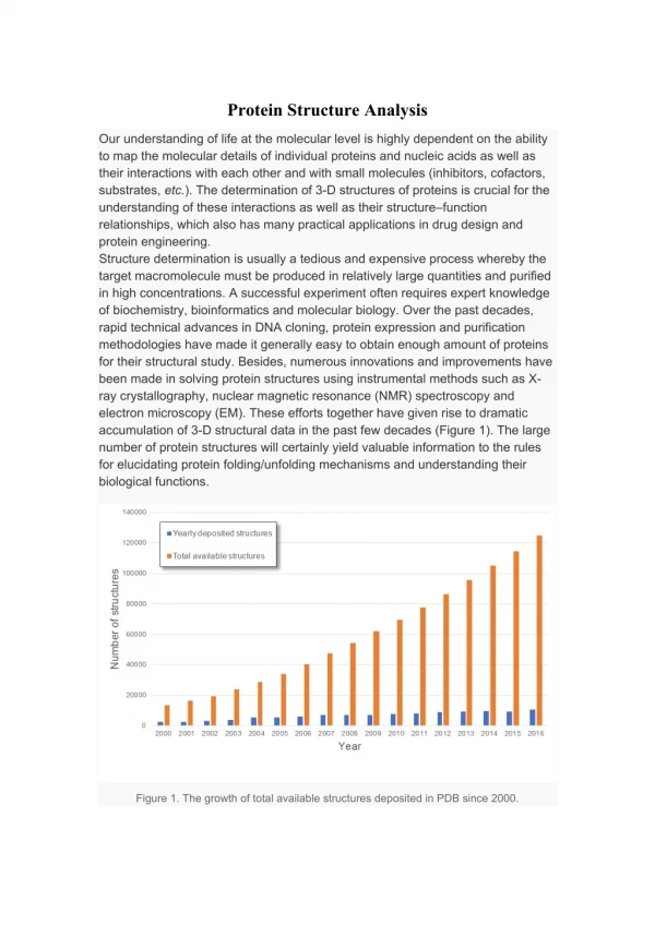
Why do we analyse proteins? • Proteins play crucial roles in nearly all biological processes. These many functions of proteins are a result of the folding of proteins into many distinct 3D structures. • Protein analysis tries to explore how amino acid sequences specify the structure of proteins and how these proteins bind to substrates and other molecules to perform their functions. • Protein analysis allows us to understand the function of the protein based on its structure.
Main Steps in Protein Analysis • Extraction of proteins. • Purification of the protein. • Structural Characterization of the protein.
1. Protein Extraction • Involves the efficient extraction of the protein and peptide in a biologically active form from tissue. • During this process, inactivation of proteolytic enzymes is required. Why ? Not to cause degradation of the proteins contained in the tissue. • Extraction of protein: homogenize it in a physiological buffer (0.05M NaPO4, PH 7.4) containing proteolytic enzyme inhibitors (EDTA, Pepstatin) • Extraction of peptides: boil the tissue with 1M Acetic Acid for 5 min homogenize tissue in ethanol / 0.1MHCl (ratio 3:1) at 0°C. This will burst open the vesicles to release the peptides in solution.
2. Protein Purification • QProteins can be purified according to certain properties they possess. These properties allow us to employ different techniques in purifying proteins.
Protein PurificationA. Differential Precipitation • Precipitation: the process of formation of a solid that was previously held in solution. The solid is separated from the solution. • When NH4 SO4 or polyethylene glycol are added to a protein solution, a precipitate forms and it can be separated from the solution after centrifugation. • If the concentrations of NH4 SO4 or polyethylene glycol are increased, more precipitate forms.
Protein PurificationB. Gel Filtration Chromatography • Also called Gel Permeation Chromatography. • Separates protein molecules according to their molecular size. • The solution is inserted to the top of a specialized column. • This column consists of specialized porous beads. • Small molecules of protein enter the beads while large molecules can’t and stay in the space between the beads. • Therefore, large molecules flow more rapidly through the column and emerge first from the bottom of the column. • Advantage: larger quantities of proteins can be separated. • Disadvantage: Lower resolution.
Protein PurificationC. Ion Exchange Chromatography • Separates protein molecules according to their molecular charge. • In this technique, the beads of the column have a specific charge on them. This is a result of a molecule that is attached to these beads. • The beads might be +ve charged by attaching them to DEAE (diethylaminoethyl) cellulose or –ve charged by attaching them to carboxymethyl cellulose. • The beads in the column depend on the protein that you want to purify. • If the protein is –ve charged then you have to use positively charged beads and vice versa.
Protein PurificationC. Ion Exchange Chromatography How does it work? • For example, if the protein of interest is negatively charged, then you will use a DEAE-cellulose column. • The protein will bind to the positively charged beads. • This protein that is attached to the beads can be released by increasing the concentration of NaCl (or other salt). • The Na+ ions (or other cation) will compete and bind to the beads in the column instead of the protein. • Proteins that are highly positively charged will emerge first because they will be repelled by the beads.
Protein PurificationD. Reversed Phase HPLC • This technique purifies proteins according to their hydrophobicity. • HPLC – High Pressure Liquid Chromatography • Reverse phase chromatography is a form of chromatography in which the stationary phase is hydrophobic and the mobile phase is more hydrophilic than the stationary phase. This is "reversed" from normal phase chromatography, in which the opposite is the case. • The protein solution is pumped into a column containing Silica beads with attached hydrocarbon chain groups. • These hydrocarbon groups may differ. Example: Octacecyl, Butyl, Propyl, Phenyldimethyl. • The hydrophobic groups of the protein will bind to the beads. • The hydrophilic proteins will emerge from the column first. • There are many different sizes of columns with varying widths: Preperative (2.5-5cm) Analytical (0.4-1cm) Microbore (0.1-0.2cm) • The solvents used: • Aqueous Phase: Water + 0.1% Triflouroacetic acid • Organic Phase: Acetonitrile
Protein PurificationE. Affinity Chromatography • It is the most powerful means of purifying proteins. • It takes the advantage of the high affinity of many proteins to specific chemical groups or molecules. • Example: Concavalin A is a glucose binding protein. It can be purified by passing it through a column that has special beads. These beads have glucose attached to them. The glucose binding protein binds to the beads and other proteins don’t. The protein can be released from the beads by increasing the concentration of glucose. This will displace the glucose binding protein from the beads.
Protein PurificationE. Affinity Chromatography • Other proteins can be separated by this method based on their affinity for specific groups or compounds. • Examples: Antigen Antibody Antibody Antigen Substrate Enzyme Concavalin A Glycoprotein Hormone Binding Protein/Receptor
3. Protein Characterization • Characterization of proteins and peptides involves three different processes: • Determining the Amino Acid Composition • Involves finding out the amino acids that make up the protein and their number. • Determining the Amino Acid Sequence • Involves finding out the sequence of amino acids of the proteins in their order. • Determining the Molecular mass of the Protein
Protein Characterization1. Determination of Amino Acid Composition • The peptide is first hydrolyzed into its constituent amino acids by heating it in 6M HCl at 110ºC for 24 hrs. This is done under vacuum or in an atmosphere of argon. • The amino acids are then separated by HPLC. • They are measured by reacting them with a compound called ninhydrin. Alpha Amino acids will be given an intense blue color while imino acids (proline, hydroproline) will be given a yellow color. • The concentration of a single amino acid is proportional to the light absorbance of the solution after adding ninhydrin. • The elution profile of the amino acids is obtained. Eluting is the separation, by washing, of one solid from another. • The identity of the amino acid is revealed by its elution volume, which is the volume of buffer needed to remove the a.a. from the column.
Protein Characterization2. Determination of Amino Acid Sequence • Pehr Edman devised a method for labeling the amino-terminal of a peptide and cleaving it from the peptide without distrupting the peptide bonds between the other a.a. residues. • This method is called Edman degradation. • It sequentially removes one residue at a time from the amino end of the peptide. • First Phenylisothiocyanate reacts with the terminal amino group to form a phenylthiocarbamoyl derivative. • This residue cyclizes under acidic conditions to give a PTH amino acid and a peptide shortened by one amino acid residue. • This PTH-amino acid is identified by HPLC. • Automated repeated Edman degradation by a sequenator that can analyze sequences of about 50 amino acids long. • The amino acid composition of the shortened peptide can be compared with the original peptide.
Protein Characterization2. Determination of Amino Acid Sequence • Longer polypeptide chains are broken into shorter ones for analysis by specifically cleaving them with enzymes that cleave at specific points. • Some examples are :
Protein Characterization3. Determination of Molecular Mass by Mass Spectrometry • Spectrometry: The procedure of observing and measuring the wavelengths of light or other electromagnetic emissions. • Mass Spectrometry involves an ionization source, a mass analyzer and a detector. The ionized hit the mass analyzer. The time the ions take in flight is recorded by triggering a laser pulse. • It depends on one principle • The smaller ions will fly faster and hit the detector first then the larger ions reach. The ions hit the detector in order by mass. • http://www.psrc.usm.edu/macrog/
Protein Characterization3. Determination of Molecular Mass by Mass Spectrometry Diagram